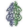
[English] 日本語
Yorodumi
- PDB-3hn3: Human beta-glucuronidase at 1.7 A resolution -

+
Open data


ID or keywords:

Loading...

-
Basic information

Entry
Database: PDB / ID: 3hn3
TitleHuman beta-glucuronidase at 1.7 A resolution
ComponentsBeta-glucuronidase
KeywordsHYDROLASE / LYSOSOMAL ENZYME / ACID HYDROLASE / GLYCOSIDASE / Disease mutation / Glycoprotein / Lysosome / Mucopolysaccharidosis
Function / homology
Function and homology information


MPS VII - Sly syndrome / chondroitin sulfate catabolic process / glycosaminoglycan catabolic process / Hyaluronan uptake and degradation / glucuronoside catabolic process / beta-glucuronidase / heparan sulfate proteoglycan catabolic process / beta-glucuronidase activity / HS-GAG degradation / hyaluronan catabolic process ...MPS VII - Sly syndrome / chondroitin sulfate catabolic process / glycosaminoglycan catabolic process / Hyaluronan uptake and degradation / glucuronoside catabolic process / beta-glucuronidase / heparan sulfate proteoglycan catabolic process / beta-glucuronidase activity / HS-GAG degradation / hyaluronan catabolic process / lysosomal lumen / azurophil granule lumen / carbohydrate binding / ficolin-1-rich granule lumen / carbohydrate metabolic process / protein domain specific binding / intracellular membrane-bounded organelle / signaling receptor binding / Neutrophil degranulation / extracellular space / extracellular exosome / extracellular region / membrane
Similarity search - Function
Glycoside hydrolase, family 2, active site / Glycosyl hydrolases family 2 acid/base catalyst. / Glycoside hydrolase, family 2, conserved site / Glycosyl hydrolases family 2 signature 1. / Glycoside hydrolase, family 2 / Glycoside hydrolase family 2, catalytic domain / Glycosyl hydrolases family 2, TIM barrel domain / Glycoside hydrolase, family 2, immunoglobulin-like beta-sandwich / Glycosyl hydrolases family 2, sugar binding domain / Glycosyl hydrolases family 2 ...Glycoside hydrolase, family 2, active site / Glycosyl hydrolases family 2 acid/base catalyst. / Glycoside hydrolase, family 2, conserved site / Glycosyl hydrolases family 2 signature 1. / Glycoside hydrolase, family 2 / Glycoside hydrolase family 2, catalytic domain / Glycosyl hydrolases family 2, TIM barrel domain / Glycoside hydrolase, family 2, immunoglobulin-like beta-sandwich / Glycosyl hydrolases family 2, sugar binding domain / Glycosyl hydrolases family 2 / Glycosyl hydrolases family 2, sugar binding domain / Beta-Galactosidase/glucuronidase domain superfamily / Galactose-binding domain-like / Galactose-binding-like domain superfamily / Glycosidases / Glycoside hydrolase superfamily / Jelly Rolls / Immunoglobulins / TIM Barrel / Alpha-Beta Barrel / Immunoglobulin-like fold / Immunoglobulin-like / Sandwich / Mainly Beta / Alpha Beta
Similarity search - Domain/homology
beta-D-mannopyranose / Beta-glucuronidase
Similarity search - Component
Biological speciesHomo sapiens (human)
MethodX-RAY DIFFRACTION / SYNCHROTRON / MOLECULAR REPLACEMENT / molecular replacement / Resolution: 1.7 Å
AuthorsKlei, H.E. / Ghosh, K. / Anumula, R.
CitationJournal: To be Published
Title: Human beta-glucuronidasel at 1.7 A resolution
Authors: Klei, H.E. / Ghosh, K. / Anumula, R.
History
DepositionMay 29, 2009Deposition site: RCSB / Processing site: PDBJ
Revision 1.0Dec 1, 2009Provider: repository / Type: Initial release
Revision 1.1Jul 13, 2011Group: Non-polymer description / Version format compliance
Revision 1.2Oct 4, 2017Group: Advisory / Data collection / Refinement description
Category: diffrn_detector / pdbx_unobs_or_zero_occ_atoms / software
Item: _diffrn_detector.detector
Revision 2.0Jul 29, 2020Group: Advisory / Atomic model ...Advisory / Atomic model / Data collection / Derived calculations / Non-polymer description / Structure summary
Category: atom_site / chem_comp ...atom_site / chem_comp / database_PDB_caveat / entity / pdbx_branch_scheme / pdbx_chem_comp_identifier / pdbx_entity_branch / pdbx_entity_branch_descriptor / pdbx_entity_branch_link / pdbx_entity_branch_list / pdbx_entity_nonpoly / pdbx_nonpoly_scheme / pdbx_struct_assembly_gen / pdbx_validate_chiral / struct_asym / struct_conn / struct_site / struct_site_gen
Item: _atom_site.B_iso_or_equiv / _atom_site.Cartn_x ..._atom_site.B_iso_or_equiv / _atom_site.Cartn_x / _atom_site.Cartn_y / _atom_site.Cartn_z / _atom_site.auth_asym_id / _atom_site.auth_atom_id / _atom_site.auth_comp_id / _atom_site.auth_seq_id / _atom_site.label_asym_id / _atom_site.label_atom_id / _atom_site.label_comp_id / _atom_site.label_entity_id / _atom_site.type_symbol / _chem_comp.formula / _chem_comp.formula_weight / _chem_comp.id / _chem_comp.mon_nstd_flag / _chem_comp.name / _chem_comp.type / _pdbx_struct_assembly_gen.asym_id_list / _struct_conn.pdbx_dist_value / _struct_conn.pdbx_leaving_atom_flag / _struct_conn.pdbx_role / _struct_conn.ptnr1_auth_asym_id / _struct_conn.ptnr1_auth_comp_id / _struct_conn.ptnr1_auth_seq_id / _struct_conn.ptnr1_label_asym_id / _struct_conn.ptnr1_label_atom_id / _struct_conn.ptnr1_label_comp_id / _struct_conn.ptnr1_label_seq_id / _struct_conn.ptnr2_auth_asym_id / _struct_conn.ptnr2_auth_comp_id / _struct_conn.ptnr2_auth_seq_id / _struct_conn.ptnr2_label_asym_id / _struct_conn.ptnr2_label_atom_id / _struct_conn.ptnr2_label_comp_id / _struct_conn.ptnr2_label_seq_id
Description: Carbohydrate remediation / Provider: repository / Type: Remediation

-
Structure visualization

Structure viewerMolecule:
MolmilJmol/JSmol

Downloads & links

-
Assembly

Deposited unit
A: Beta-glucuronidase
B: Beta-glucuronidase
D: Beta-glucuronidase
E: Beta-glucuronidase
hetero molecules


Theoretical massNumber of molelcules
Total (without water)291,59318
Polymers283,0514
Non-polymers8,54214
Water50,5682807
1


  • Idetical with deposited unit
  • defined by author&software
TypeNameSymmetry operationNumber
identity operation1_555x,y,z1
Buried area31630 Å2
ΔGint56 kcal/mol
Surface area83950 Å2
MethodPISA
Unit cell
Length a, b, c (Å)93.578, 123.115, 266.135
Angle α, β, γ (deg.)90.000, 90.000, 90.000
Int Tables number19
Space group name H-MP212121

-
Components

-
Protein , 1 types, 4 molecules ABDE

#1: Protein
Beta-glucuronidase / Beta-G1


Mass: 70762.867 Da / Num. of mol.: 4
Source method: isolated from a genetically manipulated source
Source: (gene. exp.) Homo sapiens (human) / Gene: PLACENTAL GUS / Plasmid: PMSXND / Production host: Mus musculus (house mouse) / References: UniProt: P08236, beta-glucuronidase

-
Sugars , 6 types, 9 molecules

#2: Polysaccharide alpha-D-mannopyranose-(1-2)-alpha-D-mannopyranose-(1-3)-[alpha-D-mannopyranose-(1-2)-alpha-D- ...alpha-D-mannopyranose-(1-2)-alpha-D-mannopyranose-(1-3)-[alpha-D-mannopyranose-(1-2)-alpha-D-mannopyranose-(1-6)]alpha-D-mannopyranose-(1-6)-[alpha-L-gulopyranose-(1-2)-alpha-D-mannopyranose-(1-3)]beta-D-mannopyranose-(1-4)-2-acetamido-2-deoxy-beta-D-glucopyranose-(1-4)-2-acetamido-2-deoxy-beta-D-glucopyranose


Type: oligosaccharide / Mass: 1721.527 Da / Num. of mol.: 1
Source method: isolated from a genetically manipulated source
DescriptorTypeProgram
DManpa1-2DManpa1-3[DManpa1-2DManpa1-6]DManpa1-6[LGulpa1-2DManpa1-3]DManpb1-4DGlcpNAcb1-4DGlcpNAcb1-Glycam Condensed SequenceGMML 1.0
WURCS=2.0/4,10,9/[a2122h-1b_1-5_2*NCC/3=O][a1122h-1b_1-5][a1122h-1a_1-5][a1121h-1a_1-5]/1-1-2-3-4-3-3-3-3-3/a4-b1_b4-c1_c3-d1_c6-f1_d2-e1_f3-g1_f6-i1_g2-h1_i2-j1WURCSPDB2Glycan 1.1.0
[]{[(4+1)][b-D-GlcpNAc]{[(4+1)][b-D-GlcpNAc]{[(4+1)][b-D-Manp]{[(3+1)][a-D-Manp]{[(2+1)][a-L-Gulp]{}}[(6+1)][a-D-Manp]{[(3+1)][a-D-Manp]{[(2+1)][a-D-Manp]{}}[(6+1)][a-D-Manp]{[(2+1)][a-D-Manp]{}}}}}}}LINUCSPDB-CARE
#3: Polysaccharide alpha-D-mannopyranose-(1-2)-alpha-D-mannopyranose-(1-3)-[alpha-D-mannopyranose-(1-2)-alpha-D- ...alpha-D-mannopyranose-(1-2)-alpha-D-mannopyranose-(1-3)-[alpha-D-mannopyranose-(1-2)-alpha-D-mannopyranose-(1-6)]alpha-D-mannopyranose-(1-6)-[beta-D-mannopyranose-(1-2)-alpha-D-mannopyranose-(1-3)]beta-D-mannopyranose-(1-4)-2-acetamido-2-deoxy-beta-D-glucopyranose-(1-4)-2-acetamido-2-deoxy-beta-D-glucopyranose


Type: oligosaccharide / Mass: 1721.527 Da / Num. of mol.: 1
Source method: isolated from a genetically manipulated source
DescriptorTypeProgram
DManpa1-2DManpa1-3[DManpa1-2DManpa1-6]DManpa1-6[DManpb1-2DManpa1-3]DManpb1-4DGlcpNAcb1-4DGlcpNAcb1-Glycam Condensed SequenceGMML 1.0
WURCS=2.0/3,10,9/[a2122h-1b_1-5_2*NCC/3=O][a1122h-1b_1-5][a1122h-1a_1-5]/1-1-2-3-2-3-3-3-3-3/a4-b1_b4-c1_c3-d1_c6-f1_d2-e1_f3-g1_f6-i1_g2-h1_i2-j1WURCSPDB2Glycan 1.1.0
[]{[(4+1)][b-D-GlcpNAc]{[(4+1)][b-D-GlcpNAc]{[(4+1)][b-D-Manp]{[(3+1)][a-D-Manp]{[(2+1)][b-D-Manp]{}}[(6+1)][a-D-Manp]{[(3+1)][a-D-Manp]{[(2+1)][a-D-Manp]{}}[(6+1)][a-D-Manp]{[(2+1)][a-D-Manp]{}}}}}}}LINUCSPDB-CARE
#4: Polysaccharide alpha-D-mannopyranose-(1-2)-alpha-D-mannopyranose-(1-3)-[alpha-D-mannopyranose-(1-2)-alpha-D- ...alpha-D-mannopyranose-(1-2)-alpha-D-mannopyranose-(1-3)-[alpha-D-mannopyranose-(1-2)-alpha-D-mannopyranose-(1-6)]alpha-D-mannopyranose-(1-6)-[alpha-D-mannopyranose-(1-2)-alpha-D-mannopyranose-(1-3)]beta-D-mannopyranose-(1-4)-2-acetamido-2-deoxy-beta-D-glucopyranose-(1-4)-2-acetamido-2-deoxy-beta-D-glucopyranose


Type: oligosaccharide / Mass: 1721.527 Da / Num. of mol.: 1
Source method: isolated from a genetically manipulated source
DescriptorTypeProgram
DManpa1-2DManpa1-3[DManpa1-2DManpa1-6]DManpa1-6[DManpa1-2DManpa1-3]DManpb1-4DGlcpNAcb1-4DGlcpNAcb1-Glycam Condensed SequenceGMML 1.0
WURCS=2.0/3,10,9/[a2122h-1b_1-5_2*NCC/3=O][a1122h-1b_1-5][a1122h-1a_1-5]/1-1-2-3-3-3-3-3-3-3/a4-b1_b4-c1_c3-d1_c6-f1_d2-e1_f3-g1_f6-i1_g2-h1_i2-j1WURCSPDB2Glycan 1.1.0
[]{[(4+1)][b-D-GlcpNAc]{[(4+1)][b-D-GlcpNAc]{[(4+1)][b-D-Manp]{[(3+1)][a-D-Manp]{[(2+1)][a-D-Manp]{}}[(6+1)][a-D-Manp]{[(3+1)][a-D-Manp]{[(2+1)][a-D-Manp]{}}[(6+1)][a-D-Manp]{[(2+1)][a-D-Manp]{}}}}}}}LINUCSPDB-CARE
#5: Polysaccharide alpha-D-mannopyranose-(1-2)-alpha-D-mannopyranose-(1-2)-alpha-D-mannopyranose-(1-3)-[alpha-D- ...alpha-D-mannopyranose-(1-2)-alpha-D-mannopyranose-(1-2)-alpha-D-mannopyranose-(1-3)-[alpha-D-mannopyranose-(1-2)-alpha-D-mannopyranose-(1-6)-[alpha-D-mannopyranose-(1-3)]alpha-D-mannopyranose-(1-6)]beta-D-mannopyranose-(1-4)-2-acetamido-2-deoxy-beta-D-glucopyranose-(1-4)-2-acetamido-2-deoxy-beta-D-glucopyranose


Type: oligosaccharide / Mass: 1721.527 Da / Num. of mol.: 1
Source method: isolated from a genetically manipulated source
DescriptorTypeProgram
DManpa1-2DManpa1-2DManpa1-3[DManpa1-2DManpa1-6[DManpa1-3]DManpa1-6]DManpb1-4DGlcpNAcb1-4DGlcpNAcb1-Glycam Condensed SequenceGMML 1.0
WURCS=2.0/3,10,9/[a2122h-1b_1-5_2*NCC/3=O][a1122h-1b_1-5][a1122h-1a_1-5]/1-1-2-3-3-3-3-3-3-3/a4-b1_b4-c1_c3-d1_c6-g1_d2-e1_e2-f1_g3-h1_g6-i1_i2-j1WURCSPDB2Glycan 1.1.0
[]{[(4+1)][b-D-GlcpNAc]{[(4+1)][b-D-GlcpNAc]{[(4+1)][b-D-Manp]{[(3+1)][a-D-Manp]{[(2+1)][a-D-Manp]{[(2+1)][a-D-Manp]{}}}[(6+1)][a-D-Manp]{[(3+1)][a-D-Manp]{}[(6+1)][a-D-Manp]{[(2+1)][a-D-Manp]{}}}}}}}LINUCSPDB-CARE
#6: Sugar
ChemComp-NAG / 2-acetamido-2-deoxy-beta-D-glucopyranose


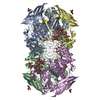
Type: D-saccharide, beta linking / Mass: 221.208 Da / Num. of mol.: 4
Source method: isolated from a genetically manipulated source
Formula: C8H15NO6
IdentifierTypeProgram
DGlcpNAcbCONDENSED IUPAC CARBOHYDRATE SYMBOLGMML 1.0
N-acetyl-b-D-glucopyranosamineCOMMON NAMEGMML 1.0
b-D-GlcpNAcIUPAC CARBOHYDRATE SYMBOLPDB-CARE 1.0
GlcNAcSNFG CARBOHYDRATE SYMBOLGMML 1.0
#9: Sugar ChemComp-BMA / beta-D-mannopyranose


Type: D-saccharide, beta linking / Mass: 180.156 Da / Num. of mol.: 1
Source method: isolated from a genetically manipulated source
Formula: C6H12O6
IdentifierTypeProgram
DManpbCONDENSED IUPAC CARBOHYDRATE SYMBOLGMML 1.0
b-D-mannopyranoseCOMMON NAMEGMML 1.0
b-D-ManpIUPAC CARBOHYDRATE SYMBOLPDB-CARE 1.0
ManSNFG CARBOHYDRATE SYMBOLGMML 1.0

-
Non-polymers , 3 types, 2812 molecules

#7: Chemical ChemComp-MRD / (4R)-2-METHYLPENTANE-2,4-DIOL


Mass: 118.174 Da / Num. of mol.: 3 / Source method: obtained synthetically / Formula: C6H14O2 / Comment: precipitant*YM
#8: Chemical ChemComp-MPD / (4S)-2-METHYL-2,4-PENTANEDIOL


Mass: 118.174 Da / Num. of mol.: 2 / Source method: obtained synthetically / Formula: C6H14O2 / Comment: precipitant*YM
#10: Water ChemComp-HOH / water


Mass: 18.015 Da / Num. of mol.: 2807 / Source method: isolated from a natural source / Formula: H2O

-
Experimental details

-
Experiment

ExperimentMethod: X-RAY DIFFRACTION / Number of used crystals: 1

-
Sample preparation

CrystalDensity Matthews: 2.71 Å3/Da / Density % sol: 54.58 %
Crystal growTemperature: 277 K / Method: vapor diffusion, hanging drop / pH: 7.5
Details: 10mM Tris-HCl pH 7.5, 15% v/v MPD, VAPOR DIFFUSION, HANGING DROP, temperature 277K

-
Data collection

DiffractionMean temperature: 100 K
Diffraction sourceSource: SYNCHROTRON / Site: APS / Beamline: 17-ID / Wavelength: 1 Å
DetectorType: Bruker AXIOM 200 / Detector: CCD / Date: Jun 19, 1998
RadiationProtocol: SINGLE WAVELENGTH / Monochromatic (M) / Laue (L): M / Scattering type: x-ray
Radiation wavelengthWavelength: 1 Å / Relative weight: 1
ReflectionResolution: 1.67→20 Å / Num. obs: 316019 / % possible obs: 88.4 % / Rmerge(I) obs: 0.103 / Χ2: 1.119 / Net I/σ(I): 11.182
Reflection shell
Resolution (Å)Rmerge(I) obsNum. unique allΧ2Diffraction-ID% possible all
1.67-1.730.488260410.321173.6
1.73-1.80.366272820.341176.8
1.8-1.880.297285060.379180.3
1.88-1.980.264298980.559184.1
1.98-2.10.183311760.626187.6
2.1-2.270.154323140.837190.6
2.27-2.490.126336211.082194.1
2.49-2.850.111345491.36196.4
2.85-3.590.087358101.981199.4
3.59-200.068368222.044199.9

-
Phasing

PhasingMethod: molecular replacement

-
Processing

Software
NameVersionClassificationNB
DENZOdata reduction
SCALEPACKdata scaling
AMoREphasing
REFMAC5.2.0019refinement
PDB_EXTRACT3.005data extraction
HKL-2000data reduction
HKL-2000data scaling
RefinementMethod to determine structure: MOLECULAR REPLACEMENT / Resolution: 1.7→19.9 Å / Cor.coef. Fo:Fc: 0.923 / Cor.coef. Fo:Fc free: 0.898 / Occupancy max: 1 / Occupancy min: 0 / SU B: 2.22 / SU ML: 0.076 / Cross valid method: THROUGHOUT / σ(F): 0 / ESU R: 0.126 / ESU R Free: 0.122 / Stereochemistry target values: MAXIMUM LIKELIHOOD / Details: HYDROGENS HAVE BEEN ADDED IN THE RIDING POSITIONS
RfactorNum. reflection% reflectionSelection details
Rfree0.24 15434 5.1 %RANDOM
Rwork0.203 ---
obs0.205 304658 90.66 %-
Solvent computationIon probe radii: 0.8 Å / Shrinkage radii: 0.8 Å / VDW probe radii: 1.2 Å / Solvent model: BABINET MODEL WITH MASK
Displacement parametersBiso max: 63.93 Å2 / Biso mean: 15.528 Å2 / Biso min: 3.84 Å2
Baniso -1Baniso -2Baniso -3
1-0.47 Å20 Å20 Å2
2--0.04 Å20 Å2
3----0.5 Å2
Refinement stepCycle: LAST / Resolution: 1.7→19.9 Å
ProteinNucleic acidLigandSolventTotal
Num. atoms19858 0 572 2807 23237
Refine LS restraints
Refine-IDTypeDev idealDev ideal targetNumber
X-RAY DIFFRACTIONr_bond_refined_d0.0090.02221189
X-RAY DIFFRACTIONr_bond_other_d0.0020.0214299
X-RAY DIFFRACTIONr_angle_refined_deg1.2641.97228940
X-RAY DIFFRACTIONr_angle_other_deg1.031334524
X-RAY DIFFRACTIONr_dihedral_angle_1_deg6.39752454
X-RAY DIFFRACTIONr_dihedral_angle_2_deg36.00723.5231036
X-RAY DIFFRACTIONr_dihedral_angle_3_deg13.42153332
X-RAY DIFFRACTIONr_dihedral_angle_4_deg17.81115140
X-RAY DIFFRACTIONr_chiral_restr0.080.23197
X-RAY DIFFRACTIONr_gen_planes_refined0.0040.0222999
X-RAY DIFFRACTIONr_gen_planes_other0.0010.024477
X-RAY DIFFRACTIONr_nbd_refined0.1970.24127
X-RAY DIFFRACTIONr_nbd_other0.1990.215652
X-RAY DIFFRACTIONr_nbtor_refined0.1820.210276
X-RAY DIFFRACTIONr_nbtor_other0.0860.210802
X-RAY DIFFRACTIONr_xyhbond_nbd_refined0.1440.22399
X-RAY DIFFRACTIONr_xyhbond_nbd_other0.0560.21
X-RAY DIFFRACTIONr_symmetry_vdw_refined0.2160.216
X-RAY DIFFRACTIONr_symmetry_vdw_other0.1950.257
X-RAY DIFFRACTIONr_symmetry_hbond_refined0.1490.251
X-RAY DIFFRACTIONr_mcbond_it0.5821.512420
X-RAY DIFFRACTIONr_mcbond_other0.1351.54898
X-RAY DIFFRACTIONr_mcangle_it0.944219790
X-RAY DIFFRACTIONr_scbond_it1.359310050
X-RAY DIFFRACTIONr_scangle_it2.0344.59136
LS refinement shellResolution: 1.7→1.744 Å / Total num. of bins used: 20
RfactorNum. reflection% reflection
Rfree0.31 976 -
Rwork0.233 18712 -
all-19688 -
obs--80.07 %

+
About Yorodumi

-
News

-
Feb 9, 2022. New format data for meta-information of EMDB entries

New format data for meta-information of EMDB entries

  • Version 3 of the EMDB header file is now the official format.
  • The previous official version 1.9 will be removed from the archive.

Related info.:EMDB header

External links:wwPDB to switch to version 3 of the EMDB data model

-
Aug 12, 2020. Covid-19 info

Covid-19 info

URL: https://pdbjlvh1.pdbj.org/emnavi/covid19.php

New page: Covid-19 featured information page in EM Navigator.

Related info.:Covid-19 info / Mar 5, 2020. Novel coronavirus structure data

+
Mar 5, 2020. Novel coronavirus structure data

Novel coronavirus structure data

Related info.:Yorodumi Speices / Aug 12, 2020. Covid-19 info

External links:COVID-19 featured content - PDBj / Molecule of the Month (242):Coronavirus Proteases

+
Jan 31, 2019. EMDB accession codes are about to change! (news from PDBe EMDB page)

EMDB accession codes are about to change! (news from PDBe EMDB page)

  • The allocation of 4 digits for EMDB accession codes will soon come to an end. Whilst these codes will remain in use, new EMDB accession codes will include an additional digit and will expand incrementally as the available range of codes is exhausted. The current 4-digit format prefixed with “EMD-” (i.e. EMD-XXXX) will advance to a 5-digit format (i.e. EMD-XXXXX), and so on. It is currently estimated that the 4-digit codes will be depleted around Spring 2019, at which point the 5-digit format will come into force.
  • The EM Navigator/Yorodumi systems omit the EMD- prefix.

Related info.:Q: What is EMD? / ID/Accession-code notation in Yorodumi/EM Navigator

External links:EMDB Accession Codes are Changing Soon! / Contact to PDBj

+
Jul 12, 2017. Major update of PDB

Major update of PDB

  • wwPDB released updated PDB data conforming to the new PDBx/mmCIF dictionary.
  • This is a major update changing the version number from 4 to 5, and with Remediation, in which all the entries are updated.
  • In this update, many items about electron microscopy experimental information are reorganized (e.g. em_software).
  • Now, EM Navigator and Yorodumi are based on the updated data.

External links:wwPDB Remediation / Enriched Model Files Conforming to OneDep Data Standards Now Available in the PDB FTP Archive

-
Yorodumi

Thousand views of thousand structures

  • Yorodumi is a browser for structure data from EMDB, PDB, SASBDB, etc.
  • This page is also the successor to EM Navigator detail page, and also detail information page/front-end page for Omokage search.
  • The word "yorodu" (or yorozu) is an old Japanese word meaning "ten thousand". "mi" (miru) is to see.

Related info.:EMDB / PDB / SASBDB / Comparison of 3 databanks / Yorodumi Search / Aug 31, 2016. New EM Navigator & Yorodumi / Yorodumi Papers / Jmol/JSmol / Function and homology information / Changes in new EM Navigator and Yorodumi

Read more