[English] 日本語
Yorodumi
- PDB-5c70: The structure of Aspergillus oryzae beta-glucuronidase -

+
Open data


ID or keywords:

Loading...

-
Basic information

Entry
Database: PDB / ID: 5c70
TitleThe structure of Aspergillus oryzae beta-glucuronidase
ComponentsGlucuronidase
KeywordsHYDROLASE / beta-glucuronidase
Function / homology
Function and homology information


glucuronoside catabolic process / beta-glucuronidase / beta-glucuronidase activity / carbohydrate binding / carbohydrate metabolic process
Similarity search - Function
Glycoside hydrolase, family 2, active site / Glycosyl hydrolases family 2 acid/base catalyst. / Glycoside hydrolase, family 2, conserved site / Glycosyl hydrolases family 2 signature 1. / Glycoside hydrolase, family 2 / Glycosyl hydrolases family 2, sugar binding domain / Glycoside hydrolase family 2, catalytic domain / Glycosyl hydrolases family 2, sugar binding domain / Glycosyl hydrolases family 2, TIM barrel domain / Glycoside hydrolase, family 2, immunoglobulin-like beta-sandwich ...Glycoside hydrolase, family 2, active site / Glycosyl hydrolases family 2 acid/base catalyst. / Glycoside hydrolase, family 2, conserved site / Glycosyl hydrolases family 2 signature 1. / Glycoside hydrolase, family 2 / Glycosyl hydrolases family 2, sugar binding domain / Glycoside hydrolase family 2, catalytic domain / Glycosyl hydrolases family 2, sugar binding domain / Glycosyl hydrolases family 2, TIM barrel domain / Glycoside hydrolase, family 2, immunoglobulin-like beta-sandwich / Glycosyl hydrolases family 2 / Beta-Galactosidase/glucuronidase domain superfamily / Galactose-binding domain-like / Galactose-binding-like domain superfamily / Glycosidases / Glycoside hydrolase superfamily / Jelly Rolls / TIM Barrel / Alpha-Beta Barrel / Immunoglobulin-like fold / Immunoglobulins / Immunoglobulin-like / Sandwich / Mainly Beta / Alpha Beta
Similarity search - Domain/homology
Biological speciesAspergillus oryzae (mold)
MethodX-RAY DIFFRACTION / SYNCHROTRON / MOLECULAR REPLACEMENT / molecular replacement / Resolution: 3.1 Å
AuthorsSun, H.L. / Lv, B. / Huang, S. / Sun, Q.F. / Li, C. / Jiang, T.
CitationJournal: Ind Eng Chem Res / Year: 2016
Title: Enhancing the Thermostability of beta-Glucuronidase by Rationally Redesigning the Catalytic Domain Based on Sequence Alignment Strategy
Authors: Feng, X.D. / Tang, H. / Han, B.J. / Lv, B. / Li, C.
History
DepositionJun 24, 2015Deposition site: RCSB / Processing site: PDBJ
Revision 1.0Jun 15, 2016Provider: repository / Type: Initial release
Revision 1.1Mar 1, 2023Group: Database references / Derived calculations / Structure summary
Category: database_2 / entity ...database_2 / entity / pdbx_struct_oper_list / struct_ref / struct_ref_seq / struct_ref_seq_dif
Item: _database_2.pdbx_DOI / _database_2.pdbx_database_accession ..._database_2.pdbx_DOI / _database_2.pdbx_database_accession / _entity.pdbx_ec / _pdbx_struct_oper_list.symmetry_operation / _struct_ref.db_code / _struct_ref.db_name / _struct_ref.pdbx_db_accession / _struct_ref.pdbx_seq_one_letter_code / _struct_ref_seq.db_align_beg / _struct_ref_seq.pdbx_auth_seq_align_beg / _struct_ref_seq.pdbx_db_accession / _struct_ref_seq.seq_align_beg
Revision 1.2May 29, 2024Group: Data collection / Category: chem_comp_atom / chem_comp_bond

-
Structure visualization

Structure viewerMolecule:
MolmilJmol/JSmol

Downloads & links

-
Assembly

Deposited unit
A: Glucuronidase
B: Glucuronidase


Theoretical massNumber of molelcules
Total (without water)136,9632
Polymers136,9632
Non-polymers00
Water00
1
A: Glucuronidase
B: Glucuronidase

A: Glucuronidase
B: Glucuronidase


Theoretical massNumber of molelcules
Total (without water)273,9254
Polymers273,9254
Non-polymers00
Water0
TypeNameSymmetry operationNumber
identity operation1_555x,y,z1
crystal symmetry operation8_555x-y,-y,-z1
Buried area14340 Å2
ΔGint-89 kcal/mol
Surface area79210 Å2
MethodPISA
Unit cell
Length a, b, c (Å)110.320, 110.320, 480.722
Angle α, β, γ (deg.)90.000, 90.000, 120.000
Int Tables number178
Space group name H-MP6122

-
Components

#1: Protein Glucuronidase


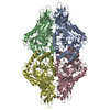
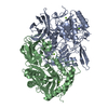
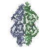
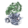
Mass: 68481.273 Da / Num. of mol.: 2
Source method: isolated from a genetically manipulated source
Source: (gene. exp.) Aspergillus oryzae (mold) / Strain: Li-3 / Gene: Pgus / Plasmid: PET28A
Production host: Escherichia coli 'BL21-Gold(DE3)pLysS AG' (bacteria)
Strain (production host): BL21-Gold(DE3)pLysS AG / References: UniProt: A7XS03, beta-mannosidase
Sequence detailsTHE SEQUENCE OF THIS PROTEIN WAS NOT AVAILABLE AT THE UNIPROT KNOWLEDGEBASE DATABASE (UNIPROTKB) AT ...THE SEQUENCE OF THIS PROTEIN WAS NOT AVAILABLE AT THE UNIPROT KNOWLEDGEBASE DATABASE (UNIPROTKB) AT THE TIME OF DEPOSITION. AUTHORS STATE THAT THE GENEBANK ACCESSION NUMBER IS EU095019.1 FOR THIS SEQUENCE.

-
Experimental details

-
Experiment

ExperimentMethod: X-RAY DIFFRACTION / Number of used crystals: 1

-
Sample preparation

CrystalDensity Matthews: 2.96 Å3/Da / Density % sol: 58.47 %
Crystal growTemperature: 289 K / Method: vapor diffusion, hanging drop / pH: 7 / Details: 1 M Succinic acid / PH range: 6.8-7.0

-
Data collection

DiffractionMean temperature: 100 K
Diffraction sourceSource: SYNCHROTRON / Site: SSRF / Beamline: BL17U / Wavelength: 1.29996 Å
DetectorType: ADSC QUANTUM 315 / Detector: CCD / Date: Jan 14, 2011
RadiationProtocol: SINGLE WAVELENGTH / Monochromatic (M) / Laue (L): M / Scattering type: x-ray
Radiation wavelengthWavelength: 1.29996 Å / Relative weight: 1
ReflectionResolution: 3.1→50 Å / Num. obs: 32783 / % possible obs: 100 % / Redundancy: 12.8 % / Rmerge(I) obs: 0.191 / Χ2: 2.141 / Net I/σ(I): 17.59 / Num. measured all: 421087
Reflection shell

Diffraction-ID: 1 / Rejects: _

Resolution (Å)Redundancy (%)Rmerge(I) obsNum. unique allΧ2% possible all
3.1-3.2112.70.47131551.39399.9
3.21-3.3413.30.39332021.529100
3.34-3.4913.10.30431761.851100
3.49-3.6813.10.25832412.073100
3.68-3.9113.20.22831982.135100
3.91-4.21130.19432322.334100
4.21-4.6312.90.17432722.50399.9
4.63-5.312.80.15932992.56999.9
5.3-6.6712.70.17333492.543100
6.67-5011.90.11836592.437100

-
Phasing

PhasingMethod: molecular replacement

-
Processing

Software
NameVersionClassification
HKL-2000data processing
HKL-2000data scaling
AMoREphasing
REFMAC5.5.0109refinement
Cootmodel building
RefinementMethod to determine structure: MOLECULAR REPLACEMENT / Resolution: 3.1→30 Å / Cor.coef. Fo:Fc: 0.925 / Cor.coef. Fo:Fc free: 0.878 / SU B: 18.567 / SU ML: 0.325 / Cross valid method: THROUGHOUT / σ(F): 0 / ESU R Free: 0.442 / Stereochemistry target values: MAXIMUM LIKELIHOOD
Details: HYDROGENS HAVE BEEN ADDED IN THE RIDING POSITIONS U VALUES : REFINED INDIVIDUALLY
RfactorNum. reflection% reflectionSelection details
Rfree0.2536 1641 5.1 %RANDOM
Rwork0.1976 ---
obs0.2003 30703 99.09 %-
Solvent computationIon probe radii: 0.8 Å / Shrinkage radii: 0.8 Å / VDW probe radii: 1.4 Å / Solvent model: MASK
Displacement parametersBiso max: 100.42 Å2 / Biso mean: 46.653 Å2 / Biso min: 23.09 Å2
Baniso -1Baniso -2Baniso -3
1-2.01 Å21.01 Å20 Å2
2--2.01 Å20 Å2
3----3.02 Å2
Refinement stepCycle: final / Resolution: 3.1→30 Å
ProteinNucleic acidLigandSolventTotal
Num. atoms9320 0 0 0 9320
Num. residues----1172
Refine LS restraints
Refine-IDTypeDev idealDev ideal targetNumber
X-RAY DIFFRACTIONr_bond_refined_d0.0110.0229560
X-RAY DIFFRACTIONr_angle_refined_deg1.4141.92613017
X-RAY DIFFRACTIONr_dihedral_angle_1_deg8.19951167
X-RAY DIFFRACTIONr_dihedral_angle_2_deg37.70723.962472
X-RAY DIFFRACTIONr_dihedral_angle_3_deg21.424151510
X-RAY DIFFRACTIONr_dihedral_angle_4_deg22.4371558
X-RAY DIFFRACTIONr_chiral_restr0.1070.21424
X-RAY DIFFRACTIONr_gen_planes_refined0.0080.0217402
X-RAY DIFFRACTIONr_mcbond_it0.6541.55828
X-RAY DIFFRACTIONr_mcangle_it1.26629418
X-RAY DIFFRACTIONr_scbond_it1.68933732
X-RAY DIFFRACTIONr_scangle_it2.9384.53599
LS refinement shellResolution: 3.101→3.181 Å / Total num. of bins used: 20
RfactorNum. reflection% reflection
Rfree0.323 123 -
Rwork0.259 2170 -
all-2293 -
obs--98.67 %

+
About Yorodumi

-
News

-
Feb 9, 2022. New format data for meta-information of EMDB entries

New format data for meta-information of EMDB entries

  • Version 3 of the EMDB header file is now the official format.
  • The previous official version 1.9 will be removed from the archive.

Related info.:EMDB header

External links:wwPDB to switch to version 3 of the EMDB data model

-
Aug 12, 2020. Covid-19 info

Covid-19 info

URL: https://pdbj.org/emnavi/covid19.php

New page: Covid-19 featured information page in EM Navigator.

Related info.:Covid-19 info / Mar 5, 2020. Novel coronavirus structure data

+
Mar 5, 2020. Novel coronavirus structure data

Novel coronavirus structure data

Related info.:Yorodumi Speices / Aug 12, 2020. Covid-19 info

External links:COVID-19 featured content - PDBj / Molecule of the Month (242):Coronavirus Proteases

+
Jan 31, 2019. EMDB accession codes are about to change! (news from PDBe EMDB page)

EMDB accession codes are about to change! (news from PDBe EMDB page)

  • The allocation of 4 digits for EMDB accession codes will soon come to an end. Whilst these codes will remain in use, new EMDB accession codes will include an additional digit and will expand incrementally as the available range of codes is exhausted. The current 4-digit format prefixed with “EMD-” (i.e. EMD-XXXX) will advance to a 5-digit format (i.e. EMD-XXXXX), and so on. It is currently estimated that the 4-digit codes will be depleted around Spring 2019, at which point the 5-digit format will come into force.
  • The EM Navigator/Yorodumi systems omit the EMD- prefix.

Related info.:Q: What is EMD? / ID/Accession-code notation in Yorodumi/EM Navigator

External links:EMDB Accession Codes are Changing Soon! / Contact to PDBj

+
Jul 12, 2017. Major update of PDB

Major update of PDB

  • wwPDB released updated PDB data conforming to the new PDBx/mmCIF dictionary.
  • This is a major update changing the version number from 4 to 5, and with Remediation, in which all the entries are updated.
  • In this update, many items about electron microscopy experimental information are reorganized (e.g. em_software).
  • Now, EM Navigator and Yorodumi are based on the updated data.

External links:wwPDB Remediation / Enriched Model Files Conforming to OneDep Data Standards Now Available in the PDB FTP Archive

-
Yorodumi

Thousand views of thousand structures

  • Yorodumi is a browser for structure data from EMDB, PDB, SASBDB, etc.
  • This page is also the successor to EM Navigator detail page, and also detail information page/front-end page for Omokage search.
  • The word "yorodu" (or yorozu) is an old Japanese word meaning "ten thousand". "mi" (miru) is to see.

Related info.:EMDB / PDB / SASBDB / Comparison of 3 databanks / Yorodumi Search / Aug 31, 2016. New EM Navigator & Yorodumi / Yorodumi Papers / Jmol/JSmol / Function and homology information / Changes in new EM Navigator and Yorodumi

Read more