 Movie
Movie Controller

[English] 日本語
 Yorodumi
- PDB-6jz2: b-glucuronidase from Ruminococcus gnavus in complex with uronic i... -
+ Open data

- Basic information
| Entry | Database: PDB / ID: 6jz2 | ||||||
|---|---|---|---|---|---|---|---|
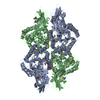
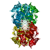
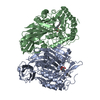
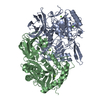
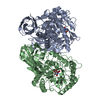
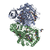
| Title | b-glucuronidase from Ruminococcus gnavus in complex with uronic isofagomine at 1.3 Angstroms resolution | ||||||
 Components Components | Beta-glucuronidase | ||||||
 Keywords Keywords | HYDROLASE / b-glucuronidase | ||||||
| Function / homology |  Function and homology information Function and homology informationglucuronoside catabolic process / beta-glucuronidase / beta-glucuronidase activity / carbohydrate binding / carbohydrate metabolic process Similarity search - Function | ||||||
| Biological species |  Ruminococcus gnavus (bacteria) Ruminococcus gnavus (bacteria) | ||||||
| Method |  X-RAY DIFFRACTION / SYNCHROTRON / MOLECULAR REPLACEMENT / Resolution: 1.29 Å X-RAY DIFFRACTION / SYNCHROTRON / MOLECULAR REPLACEMENT / Resolution: 1.29 Å | ||||||
 Authors Authors | Dashnyam, P. / Lin, H.Y. | ||||||
| Funding support |  Taiwan, 1items Taiwan, 1items
| ||||||
 Citation Citation | Journal: J.Med.Chem. / Year: 2020 Title: Substituent Position of Iminocyclitols Determines the Potency and Selectivity for Gut Microbial Xenobiotic-Reactivating Enzymes. Authors: Dashnyam, P. / Lin, H.Y. / Chen, C.Y. / Gao, S. / Yeh, L.F. / Hsieh, W.C. / Tu, Z. / Lin, C.H. | ||||||
| History |
|
- Structure visualization
| Structure viewer | Molecule: MolmilJmol/JSmol |
|---|
- Downloads & links
-Download
| PDBx/mmCIF format | 6jz2.cif.gz | 502.5 KB | Display | PDBx/mmCIF format |
|---|---|---|---|---|
| PDB format | pdb6jz2.ent.gz | 407.4 KB | Display | PDB format |
| PDBx/mmJSON format | 6jz2.json.gz | Tree view | PDBx/mmJSON format | |
| Others |  Other downloads Other downloads |
-Validation report
| Arichive directory | https://data.pdbj.org/pub/pdb/validation_reports/jz/6jz2ftp://data.pdbj.org/pub/pdb/validation_reports/jz/6jz2 | HTTPS FTP |
|---|
-Related structure data
| Related structure data |  6jz1C  6jz3C  6jz4C  6jz5C  6jz6C 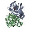 6jz7C 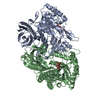 6jz8C  5z18S S: Starting model for refinement C: citing same article ( |
|---|---|
| Similar structure data |


-Links
 PDBj
PDBj

- Assembly
| Deposited unit | 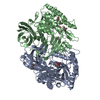
| |||||||||
|---|---|---|---|---|---|---|---|---|---|---|
| 1 | 
| |||||||||
| Unit cell |
| |||||||||
| Components on special symmetry positions |
|
-Components
| #1: Protein | Mass: 72768.805 Da / Num. of mol.: 2 Source method: isolated from a genetically manipulated source Source: (gene. exp.) Ruminococcus gnavus (bacteria) / Gene: uidA / Production host: #2: Chemical |   Mass: 161.156 Da / Num. of mol.: 2 / Source method: obtained synthetically / Formula: C6H11NO4 / Feature type: SUBJECT OF INVESTIGATION Mass: 161.156 Da / Num. of mol.: 2 / Source method: obtained synthetically / Formula: C6H11NO4 / Feature type: SUBJECT OF INVESTIGATION#3: Chemical |   Mass: 118.174 Da / Num. of mol.: 3 / Source method: obtained synthetically / Formula: C6H14O2 / Comment: precipitant*YM Mass: 118.174 Da / Num. of mol.: 3 / Source method: obtained synthetically / Formula: C6H14O2 / Comment: precipitant*YM#4: Chemical |   Mass: 118.174 Da / Num. of mol.: 2 / Source method: obtained synthetically / Formula: C6H14O2 / Comment: precipitant*YM Mass: 118.174 Da / Num. of mol.: 2 / Source method: obtained synthetically / Formula: C6H14O2 / Comment: precipitant*YM#5: Water | ChemComp-HOH / |  Mass: 18.015 Da / Num. of mol.: 1111 / Source method: isolated from a natural source / Formula: H2O Mass: 18.015 Da / Num. of mol.: 1111 / Source method: isolated from a natural source / Formula: H2OHas ligand of interest | Y | |
|---|
-Experimental details
-Experiment
| Experiment | Method: X-RAY DIFFRACTION / Number of used crystals: 1 |
|---|
- Sample preparation
| Crystal | Density Matthews: 2.55 Å3/Da / Density % sol: 51.73 % |
|---|---|
| Crystal grow | Temperature: 298 K / Method: vapor diffusion, sitting drop / Details: 70% MPD, 0.1 M HEPES pH 7.5, 0.2 M CaCl2 |
-Data collection
| Diffraction | Mean temperature: 277 K / Serial crystal experiment: N | |||||||||||||||||||||||||||||||||||||||||||||||||||||||||||||||||||||||||||||||||||||||||||||||||||
|---|---|---|---|---|---|---|---|---|---|---|---|---|---|---|---|---|---|---|---|---|---|---|---|---|---|---|---|---|---|---|---|---|---|---|---|---|---|---|---|---|---|---|---|---|---|---|---|---|---|---|---|---|---|---|---|---|---|---|---|---|---|---|---|---|---|---|---|---|---|---|---|---|---|---|---|---|---|---|---|---|---|---|---|---|---|---|---|---|---|---|---|---|---|---|---|---|---|---|---|---|
| Diffraction source | Source: SYNCHROTRON / Site: NSRRC / Beamline: BL13C1 / Wavelength: 1 Å | |||||||||||||||||||||||||||||||||||||||||||||||||||||||||||||||||||||||||||||||||||||||||||||||||||
| Detector | Type: ADSC QUANTUM 210 / Detector: CCD / Date: Oct 16, 2018 | |||||||||||||||||||||||||||||||||||||||||||||||||||||||||||||||||||||||||||||||||||||||||||||||||||
| Radiation | Protocol: SINGLE WAVELENGTH / Monochromatic (M) / Laue (L): M / Scattering type: x-ray | |||||||||||||||||||||||||||||||||||||||||||||||||||||||||||||||||||||||||||||||||||||||||||||||||||
| Radiation wavelength | Wavelength: 1 Å / Relative weight: 1 | |||||||||||||||||||||||||||||||||||||||||||||||||||||||||||||||||||||||||||||||||||||||||||||||||||
| Reflection | Resolution: 1.286→30 Å / Num. obs: 66391 / % possible obs: 95.6 % / Redundancy: 5.1 % / Rmerge(I) obs: 0.045 / Rpim(I) all: 0.022 / Rrim(I) all: 0.05 / Χ2: 0.937 / Net I/σ(I): 13 | |||||||||||||||||||||||||||||||||||||||||||||||||||||||||||||||||||||||||||||||||||||||||||||||||||
| Reflection shell | Diffraction-ID: 1
|
- Processing
| Software |
| ||||||||||||||||||||||||||||||||||||||||||||||||||||||||||||||||||||||||||||||||||||||||||
|---|---|---|---|---|---|---|---|---|---|---|---|---|---|---|---|---|---|---|---|---|---|---|---|---|---|---|---|---|---|---|---|---|---|---|---|---|---|---|---|---|---|---|---|---|---|---|---|---|---|---|---|---|---|---|---|---|---|---|---|---|---|---|---|---|---|---|---|---|---|---|---|---|---|---|---|---|---|---|---|---|---|---|---|---|---|---|---|---|---|---|---|
| Refinement | Method to determine structure: MOLECULAR REPLACEMENT Starting model: 5Z18 Resolution: 1.29→27.094 Å / SU ML: 0.11 / Cross valid method: THROUGHOUT / σ(F): 1.34 / Phase error: 17.41
| ||||||||||||||||||||||||||||||||||||||||||||||||||||||||||||||||||||||||||||||||||||||||||
| Solvent computation | Shrinkage radii: 0.9 Å / VDW probe radii: 1.11 Å | ||||||||||||||||||||||||||||||||||||||||||||||||||||||||||||||||||||||||||||||||||||||||||
| Displacement parameters | Biso max: 137.54 Å2 / Biso mean: 20.4107 Å2 / Biso min: 8.12 Å2 | ||||||||||||||||||||||||||||||||||||||||||||||||||||||||||||||||||||||||||||||||||||||||||
| Refinement step | Cycle: final / Resolution: 1.29→27.094 Å
| ||||||||||||||||||||||||||||||||||||||||||||||||||||||||||||||||||||||||||||||||||||||||||
| Refine LS restraints |
| ||||||||||||||||||||||||||||||||||||||||||||||||||||||||||||||||||||||||||||||||||||||||||
| LS refinement shell | Refine-ID: X-RAY DIFFRACTION / Rfactor Rfree error: 0
| ||||||||||||||||||||||||||||||||||||||||||||||||||||||||||||||||||||||||||||||||||||||||||
| Refinement TLS params. | Method: refined / Origin x: -54.9105 Å / Origin y: -1.75 Å / Origin z: 24.0413 Å
| ||||||||||||||||||||||||||||||||||||||||||||||||||||||||||||||||||||||||||||||||||||||||||
| Refinement TLS group |
|
