- PDB-3hfc: A trimeric form of the Kv7.1 A domain Tail, L602M/L606M mutant Semet -
+
Open data
ID or keywords:
Loading...
-
Basic information
Entry
Database: PDB / ID: 3hfc
Title
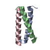
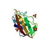
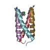
A trimeric form of the Kv7.1 A domain Tail, L602M/L606M mutant Semet
Components
Potassium voltage-gated channel subfamily KQT member 1
Keywords
TRANSPORT PROTEIN / coiled coil / trimer / Alternative splicing / Atrial fibrillation / Cell membrane / Cytoplasmic vesicle / Deafness / Disease mutation / Glycoprotein / Ion transport / Ionic channel / Long QT syndrome / Membrane / Phosphoprotein / Polymorphism / Potassium / Potassium channel / Potassium transport / Short QT syndrome / Transmembrane / Transport / Voltage-gated channel
Function / homology
Function and homology information
gastrin-induced gastric acid secretion / stomach development / corticosterone secretion / voltage-gated potassium channel activity involved in atrial cardiac muscle cell action potential repolarization / basolateral part of cell / lumenal side of membrane / negative regulation of delayed rectifier potassium channel activity / rhythmic behavior / negative regulation of voltage-gated potassium channel activity / regulation of gastric acid secretion ...gastrin-induced gastric acid secretion / stomach development / corticosterone secretion / voltage-gated potassium channel activity involved in atrial cardiac muscle cell action potential repolarization / basolateral part of cell / lumenal side of membrane / negative regulation of delayed rectifier potassium channel activity / rhythmic behavior / negative regulation of voltage-gated potassium channel activity / regulation of gastric acid secretion / voltage-gated potassium channel activity involved in cardiac muscle cell action potential repolarization / Phase 3 - rapid repolarisation / membrane repolarization during action potential / auditory receptor cell development / membrane repolarization during atrial cardiac muscle cell action potential / iodide transport / Phase 2 - plateau phase / intracellular chloride ion homeostasis / regulation of atrial cardiac muscle cell membrane repolarization / membrane repolarization during ventricular cardiac muscle cell action potential / atrial cardiac muscle cell action potential / renal sodium ion absorption / potassium ion export across plasma membrane / voltage-gated potassium channel activity involved in ventricular cardiac muscle cell action potential repolarization / regulation of membrane repolarization / membrane repolarization during cardiac muscle cell action potential / detection of mechanical stimulus involved in sensory perception of sound / protein phosphatase 1 binding / potassium ion homeostasis / intestinal absorption / delayed rectifier potassium channel activity / ventricular cardiac muscle cell action potential / regulation of ventricular cardiac muscle cell membrane repolarization / non-motile cilium assembly / positive regulation of potassium ion transmembrane transport / inner ear morphogenesis / Voltage gated Potassium channels / outward rectifier potassium channel activity / monoatomic ion channel complex / regulation of heart contraction / neuromuscular process / cardiac muscle cell contraction / adrenergic receptor signaling pathway / inner ear development / renal absorption / protein kinase A regulatory subunit binding / ciliary base / protein kinase A catalytic subunit binding / potassium ion import across plasma membrane / regulation of heart rate by cardiac conduction / social behavior / cochlea development / action potential / voltage-gated potassium channel activity / positive regulation of heart rate / cardiac muscle contraction / transport vesicle / sensory perception of sound / phosphatidylinositol-4,5-bisphosphate binding / positive regulation of cardiac muscle contraction / cellular response to epinephrine stimulus / voltage-gated potassium channel complex / cellular response to cAMP / potassium ion transmembrane transport / erythrocyte differentiation / glucose metabolic process / response to insulin / cellular response to xenobiotic stimulus / cytoplasmic vesicle membrane / regulation of membrane potential / regulation of blood pressure / heart development / late endosome / scaffold protein binding / calmodulin binding / basolateral plasma membrane / early endosome / transmembrane transporter binding / lysosome / neuron projection / apical plasma membrane / membrane raft / ubiquitin protein ligase binding / neuronal cell body / endoplasmic reticulum / membrane / plasma membrane / cytoplasm Similarity search - Function
Potassium channel, voltage dependent, KCNQ1 / Potassium channel, voltage dependent, KCNQ / Potassium channel, voltage dependent, KCNQ, C-terminal / KCNQ voltage-gated potassium channel / Voltage-dependent channel domain superfamily / Ion transport domain / Ion transport protein Similarity search - Domain/homology
Journal: Protein Sci. / Year: 2009 Title: Crystal structure of a trimeric form of the K(V)7.1 (KCNQ1) A-domain tail coiled-coil reveals structural plasticity and context dependent changes in a putative coiled-coil trimerization motif. Authors: Xu, Q. / Minor, D.L.
Resolution: 2.45→2.54 Å / Redundancy: 4.6 % / Mean I/σ(I) obs: 5.4 / Num. unique all: 238 / Rsym value: 0.203 / % possible all: 93
-
Processing
Software
Name
Version
Classification
NB
REFMAC
5.2.0005
refinement
PDB_EXTRACT
3.005
dataextraction
ADSC
Quantum
datacollection
HKL-2000
datareduction
HKL-2000
datascaling
SOLVE
phasing
Refinement
Method to determine structure: SAD / Resolution: 2.45→32.48 Å / Cor.coef. Fo:Fc: 0.928 / Cor.coef. Fo:Fc free: 0.862 / Occupancy max: 1 / Occupancy min: 1 / SU B: 9.153 / SU ML: 0.199 / Cross valid method: THROUGHOUT / σ(F): 0 / ESU R Free: 0.344 Stereochemistry target values: MAXIMUM LIKELIHOOD WITH PHASES Details: HYDROGENS HAVE BEEN ADDED IN THE RIDING POSITIONS
Rfactor
Num. reflection
% reflection
Selection details
Rfree
0.269
120
4.6 %
RANDOM
Rwork
0.221
-
-
-
obs
0.223
2600
92.43 %
-
Solvent computation
Ion probe radii: 0.8 Å / Shrinkage radii: 0.8 Å / VDW probe radii: 1.2 Å / Solvent model: MASK
In the structure databanks used in Yorodumi, some data are registered as the other names, "COVID-19 virus" and "2019-nCoV". Here are the details of the virus and the list of structure data.
Jan 31, 2019. EMDB accession codes are about to change! (news from PDBe EMDB page)
EMDB accession codes are about to change! (news from PDBe EMDB page)
The allocation of 4 digits for EMDB accession codes will soon come to an end. Whilst these codes will remain in use, new EMDB accession codes will include an additional digit and will expand incrementally as the available range of codes is exhausted. The current 4-digit format prefixed with “EMD-” (i.e. EMD-XXXX) will advance to a 5-digit format (i.e. EMD-XXXXX), and so on. It is currently estimated that the 4-digit codes will be depleted around Spring 2019, at which point the 5-digit format will come into force.
The EM Navigator/Yorodumi systems omit the EMD- prefix.
Related info.:Q: What is EMD? / ID/Accession-code notation in Yorodumi/EM Navigator
Yorodumi is a browser for structure data from EMDB, PDB, SASBDB, etc.
This page is also the successor to EM Navigator detail page, and also detail information page/front-end page for Omokage search.
The word "yorodu" (or yorozu) is an old Japanese word meaning "ten thousand". "mi" (miru) is to see.
Related info.:EMDB / PDB / SASBDB / Comparison of 3 databanks / Yorodumi Search / Aug 31, 2016. New EM Navigator & Yorodumi / Yorodumi Papers / Jmol/JSmol / Function and homology information / Changes in new EM Navigator and Yorodumi
 Movie
Movie Controller

 Yorodumi
 Open data
 Basic information
 Components
 Keywords
 Function and homology information
 Homo sapiens (human)
 X-RAY DIFFRACTION /
  Authors
 Citation
 Structure visualization
 Downloads & links
 Other downloads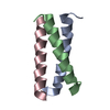
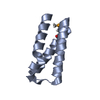
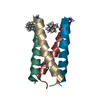
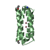
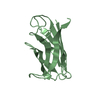
 PDBj
PDBj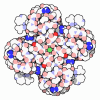
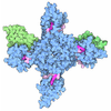
 Assembly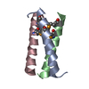

 Escherichia coli (E. coli) / Strain (production host): BL21(pLys)S / References: UniProt: P51787
 Mass: 18.015 Da / Num. of mol.: 12 / Source method: isolated from a natural source / Formula: H2O
 Sample preparation
 / Beamline: 8.3.1 / Wavelength: 0.9796 Å
 Processing