 Movie
Movie Controller


+ Open data

- Basic information
| Entry | Database: PDB / ID: 3hbm | ||||||
|---|---|---|---|---|---|---|---|
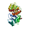
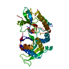
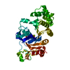
| Title | Crystal Structure of PseG from Campylobacter jejuni | ||||||
 Components Components | UDP-sugar hydrolase | ||||||
 Keywords Keywords | HYDROLASE / udp-sugar hydrolase / PseG | ||||||
| Function / homology | Rossmann fold - #11190 / UDP-2,4-diacetamido-2,4,6-trideoxy-beta-L-altropyranose hydrolase / UDP-2,4-diacetamido-2,4,6-trideoxy-beta-L-altropyranose hydrolase / Glycogen Phosphorylase B; / hydrolase activity / Rossmann fold / 3-Layer(aba) Sandwich / Alpha Beta / UDP-2,4-diacetamido-2,4,6-trideoxy-beta-L-altropyranose hydrolase Function and homology information Function and homology information | ||||||
| Biological species |  Campylobacter jejuni subsp. jejuni (Campylobacter) Campylobacter jejuni subsp. jejuni (Campylobacter) | ||||||
| Method |  X-RAY DIFFRACTION / SYNCHROTRON / SAD / Resolution: 1.8 Å X-RAY DIFFRACTION / SYNCHROTRON / SAD / Resolution: 1.8 Å | ||||||
 Authors Authors | Rangarajan, E.S. / Proteau, A. / Cygler, M. / Matte, A. / Sulea, T. / Schoenhofen, I.C. | ||||||
 Citation Citation | Journal: J.Biol.Chem. / Year: 2009 Title: Structural and functional analysis of Campylobacter jejuni PseG: a udp-sugar hydrolase from the pseudaminic acid biosynthetic pathway. Authors: Rangarajan, E.S. / Proteau, A. / Cui, Q. / Logan, S.M. / Potetinova, Z. / Whitfield, D. / Purisima, E.O. / Cygler, M. / Matte, A. / Sulea, T. / Schoenhofen, I.C. | ||||||
| History |
|
- Structure visualization
| Structure viewer | Molecule: MolmilJmol/JSmol |
|---|
- Downloads & links
-Download
| PDBx/mmCIF format | 3hbm.cif.gz | 75.4 KB | Display | PDBx/mmCIF format |
|---|---|---|---|---|
| PDB format | pdb3hbm.ent.gz | 55.9 KB | Display | PDB format |
| PDBx/mmJSON format | 3hbm.json.gz | Tree view | PDBx/mmJSON format | |
| Others |  Other downloads Other downloads |
-Validation report
| Arichive directory | https://data.pdbj.org/pub/pdb/validation_reports/hb/3hbmftp://data.pdbj.org/pub/pdb/validation_reports/hb/3hbm | HTTPS FTP |
|---|
-Related structure data


-Links
 PDBj
PDBj- Assembly
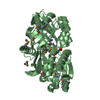
| Deposited unit | 
| ||||||||
|---|---|---|---|---|---|---|---|---|---|
| 1 |
| ||||||||
| Unit cell |
| ||||||||
| Details | Monomer |
-Components
| #1: Protein | Mass: 32572.018 Da / Num. of mol.: 1 / Mutation: E155K Source method: isolated from a genetically manipulated source Details: C-term Histag (LEHHHHHH) Source: (gene. exp.) Campylobacter jejuni subsp. jejuni (Campylobacter)Strain: NCTC 11168 / Gene: Cj1312 / Plasmid: pET30a / Production host: |
|---|---|
| #2: Chemical | ChemComp-SO4 /   Mass: 96.063 Da / Num. of mol.: 1 / Source method: obtained synthetically / Formula: SO4 Mass: 96.063 Da / Num. of mol.: 1 / Source method: obtained synthetically / Formula: SO4 |
| #3: Water | ChemComp-HOH /  Mass: 18.015 Da / Num. of mol.: 276 / Source method: isolated from a natural source / Formula: H2O Mass: 18.015 Da / Num. of mol.: 276 / Source method: isolated from a natural source / Formula: H2O |
| Has protein modification | Y |
-Experimental details
-Experiment
| Experiment | Method: X-RAY DIFFRACTION / Number of used crystals: 1 |
|---|
- Sample preparation
| Crystal | Density Matthews: 2.87 Å3/Da / Density % sol: 57.21 % |
|---|---|
| Crystal grow | Temperature: 293 K / Method: vapor diffusion, hanging drop / pH: 6.5 Details: 0.1M Bis-Tris, 0.2M Ammonium sulfate, 23% (v/v) PEG monomethyl ether 550, pH 6.5, VAPOR DIFFUSION, HANGING DROP, temperature 293K |
-Data collection
| Diffraction | Mean temperature: 100 K |
|---|---|
| Diffraction source | Source: SYNCHROTRON / Site: NSLS  / Beamline: X12B / Wavelength: 0.9791 Å / Beamline: X12B / Wavelength: 0.9791 Å |
| Detector | Type: ADSC QUANTUM 4 / Detector: CCD / Date: Apr 28, 2006 |
| Radiation | Monochromator: silicon / Protocol: SINGLE WAVELENGTH / Monochromatic (M) / Laue (L): M / Scattering type: x-ray |
| Radiation wavelength | Wavelength: 0.9791 Å / Relative weight: 1 |
| Reflection | Resolution: 1.8→33.11 Å / Num. obs: 34208 / % possible obs: 98.8 % / Observed criterion σ(F): 2 / Observed criterion σ(I): 1 / Redundancy: 7.4 % / Biso Wilson estimate: 31.4 Å2 / Rsym value: 0.073 / Net I/σ(I): 22.6 |
| Reflection shell | Resolution: 1.8→1.87 Å / Redundancy: 2 % / Mean I/σ(I) obs: 8.4 / Num. unique all: 3377 / Rsym value: 0.114 / % possible all: 97.7 |
- Processing
| Software |
| |||||||||||||||||||||||||||||||||||||||||||||||||||||||||||||||||
|---|---|---|---|---|---|---|---|---|---|---|---|---|---|---|---|---|---|---|---|---|---|---|---|---|---|---|---|---|---|---|---|---|---|---|---|---|---|---|---|---|---|---|---|---|---|---|---|---|---|---|---|---|---|---|---|---|---|---|---|---|---|---|---|---|---|---|
| Refinement | Method to determine structure: SAD / Resolution: 1.8→33.11 Å / Cor.coef. Fo:Fc: 0.951 / Cor.coef. Fo:Fc free: 0.929 / SU B: 2.485 / SU ML: 0.079 / Cross valid method: THROUGHOUT / σ(F): 0 / σ(I): 0 / ESU R: 0.122 / ESU R Free: 0.121 / Stereochemistry target values: MAXIMUM LIKELIHOOD
| |||||||||||||||||||||||||||||||||||||||||||||||||||||||||||||||||
| Solvent computation | Ion probe radii: 0.8 Å / Shrinkage radii: 0.8 Å / VDW probe radii: 1.4 Å / Solvent model: BABINET MODEL WITH MASK | |||||||||||||||||||||||||||||||||||||||||||||||||||||||||||||||||
| Displacement parameters | Biso mean: 24.736 Å2
| |||||||||||||||||||||||||||||||||||||||||||||||||||||||||||||||||
| Refinement step | Cycle: LAST / Resolution: 1.8→33.11 Å
| |||||||||||||||||||||||||||||||||||||||||||||||||||||||||||||||||
| Refine LS restraints |
| |||||||||||||||||||||||||||||||||||||||||||||||||||||||||||||||||
| LS refinement shell | Resolution: 1.801→1.848 Å / Total num. of bins used: 20
|
