Mass: 18.015 Da / Num. of mol.: 299 / Source method: isolated from a natural source / Formula: H2O
Compound details
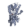
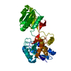
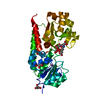
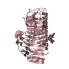
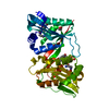
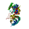
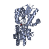
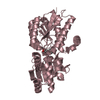
A COMPONENT OF THE LEUCINE-SPECIFIC TRANSPORT SYSTEM, WHICH IS ONE OF TWO PERIPLASMIC BINDING ...A COMPONENT OF THE LEUCINE-SPECIFIC TRANSPORT SYSTEM, WHICH IS ONE OF TWO PERIPLASMIC BINDING PROTEIN-DEPENDENT TRANSPORT SYSTEMS OF THE HIGH-AFFINITY TRANSPORT OF THE BRANCHED-CHAIN AMINO ACIDS IN E.COLI.
Has protein modification
Y
-
Experimental details
-
Experiment
Experiment
Method: X-RAY DIFFRACTION / Number of used crystals: 1
Resolution: 1.8→35 Å / Cor.coef. Fo:Fc: 0.955 / Cor.coef. Fo:Fc free: 0.936 / SU B: 2.849 / SU ML: 0.086 / Cross valid method: THROUGHOUT / ESU R: 0.132 / ESU R Free: 0.124 / Stereochemistry target values: MAXIMUM LIKELIHOOD / Details: HYDROGENS HAVE BEEN ADDED IN THE RIDING POSITIONS.
Rfactor
Num. reflection
% reflection
Selection details
Rfree
0.222
3242
5.1 %
RANDOM
Rwork
0.189
-
-
-
obs
0.191
60887
96.2 %
-
Solvent computation
Ion probe radii: 0.8 Å / Shrinkage radii: 0.8 Å / VDW probe radii: 1.4 Å / Solvent model: BABINET MODEL WITH MASK
 Movie
Movie Controller


 Open data
 Basic information
 Components
 Keywords
 Function and homology information
 X-RAY DIFFRACTION /
  Authors
 Citation
 Structure visualization
 Downloads & links
 Other downloads


 PDBj
PDBj


 Assembly
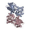

 Type: L-peptide linking / Mass: 165.189 Da / Num. of mol.: 2 / Source method: obtained synthetically / Formula: C9H11NO2
 Mass: 18.015 Da / Num. of mol.: 299 / Source method: isolated from a natural source / Formula: H2O
 Sample preparation
 / Beamline: I711 / Wavelength: 0.98
  Processing