- PDB-3h9y: Crystal structure of the IgE-Fc3-4 domains -
+
Open data
ID or keywords:
Loading...
-
Basic information
Entry
Database: PDB / ID: 3h9y
Title
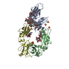
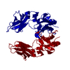
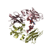
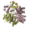
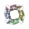
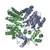
Crystal structure of the IgE-Fc3-4 domains
Components
Ig epsilon chain C region
Keywords
IMMUNE SYSTEM / Immunoglobin / IgE / Fc / flexibility / hydrophobic pocket / Disulfide bond / Glycoprotein / Immunoglobulin C region / Immunoglobulin domain
Function / homology
Function and homology information
mononuclear cell differentiation / IgE B cell receptor complex / adaptive immune memory response / primary adaptive immune response / B cell antigen processing and presentation / type I hypersensitivity / Fc receptor-mediated immune complex endocytosis / eosinophil degranulation / IgE immunoglobulin complex / macrophage activation ...mononuclear cell differentiation / IgE B cell receptor complex / adaptive immune memory response / primary adaptive immune response / B cell antigen processing and presentation / type I hypersensitivity / Fc receptor-mediated immune complex endocytosis / eosinophil degranulation / IgE immunoglobulin complex / macrophage activation / antibody-dependent cellular cytotoxicity / Fc epsilon receptor (FCERI) signaling / type 2 immune response / immunoglobulin complex, circulating / immunoglobulin receptor binding / mast cell degranulation / B cell proliferation / macrophage differentiation / B cell activation / Role of LAT2/NTAL/LAB on calcium mobilization / complement activation, classical pathway / antigen binding / FCERI mediated Ca+2 mobilization / B cell receptor signaling pathway / FCERI mediated MAPK activation / FCERI mediated NF-kB activation / antibacterial humoral response / Interleukin-4 and Interleukin-13 signaling / adaptive immune response / immune response / inflammatory response / : / extracellular region / plasma membrane Similarity search - Function
Mass: 18.015 Da / Num. of mol.: 247 / Source method: isolated from a natural source / Formula: H2O
Has protein modification
Y
-
Experimental details
-
Experiment
Experiment
Method: X-RAY DIFFRACTION / Number of used crystals: 1
-
Sample preparation
Crystal
Density Matthews: 2.6 Å3/Da / Density % sol: 52.72 %
Crystal grow
Temperature: 293 K / Method: vapor diffusion, hanging drop / pH: 5.3 Details: 1 microliter of protein at 10 mg/mL in 20 mM sodium chloride was added to 1 microliter well solution (100 mM ammonium acetate, 100 mM sodium acetate pH 4.6, 30% (w/v) PEG 4000) and mixed by ...Details: 1 microliter of protein at 10 mg/mL in 20 mM sodium chloride was added to 1 microliter well solution (100 mM ammonium acetate, 100 mM sodium acetate pH 4.6, 30% (w/v) PEG 4000) and mixed by pipetting., VAPOR DIFFUSION, HANGING DROP, temperature 293K
In the structure databanks used in Yorodumi, some data are registered as the other names, "COVID-19 virus" and "2019-nCoV". Here are the details of the virus and the list of structure data.
Jan 31, 2019. EMDB accession codes are about to change! (news from PDBe EMDB page)
EMDB accession codes are about to change! (news from PDBe EMDB page)
The allocation of 4 digits for EMDB accession codes will soon come to an end. Whilst these codes will remain in use, new EMDB accession codes will include an additional digit and will expand incrementally as the available range of codes is exhausted. The current 4-digit format prefixed with “EMD-” (i.e. EMD-XXXX) will advance to a 5-digit format (i.e. EMD-XXXXX), and so on. It is currently estimated that the 4-digit codes will be depleted around Spring 2019, at which point the 5-digit format will come into force.
The EM Navigator/Yorodumi systems omit the EMD- prefix.
Related info.:Q: What is EMD? / ID/Accession-code notation in Yorodumi/EM Navigator
Yorodumi is a browser for structure data from EMDB, PDB, SASBDB, etc.
This page is also the successor to EM Navigator detail page, and also detail information page/front-end page for Omokage search.
The word "yorodu" (or yorozu) is an old Japanese word meaning "ten thousand". "mi" (miru) is to see.
Related info.:EMDB / PDB / SASBDB / Comparison of 3 databanks / Yorodumi Search / Aug 31, 2016. New EM Navigator & Yorodumi / Yorodumi Papers / Jmol/JSmol / Function and homology information / Changes in new EM Navigator and Yorodumi
 Movie
Movie Controller


 Open data
 Basic information
 Components
 Keywords
 Function and homology information
 Homo sapiens (human)
 X-RAY DIFFRACTION /
  Authors
 Citation
 Structure visualization
 Downloads & links
 Other downloads
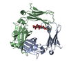


 PDBj
PDBj


 Assembly
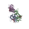


 Trichoplusia ni (cabbage looper) / Strain (production host): Hi5 / References: UniProt: P01854
 Mass: 18.038 Da / Num. of mol.: 1 / Source method: obtained synthetically / Formula: H4N
 Mass: 18.015 Da / Num. of mol.: 247 / Source method: isolated from a natural source / Formula: H2O
 Sample preparation
 / Beamline: 5ID-B / Wavelength: 1.00796 Å
 Processing