 Movie
Movie Controller

[English] 日本語
 Yorodumi
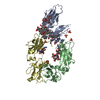
- PDB-1fp5: CRYSTAL STRUCTURE ANALYSIS OF THE HUMAN IGE-FC CEPSILON3-CEPSILON... -
+ Open data

- Basic information
| Entry | Database: PDB / ID: 1fp5 | ||||||
|---|---|---|---|---|---|---|---|
| Title | CRYSTAL STRUCTURE ANALYSIS OF THE HUMAN IGE-FC CEPSILON3-CEPSILON4 FRAGMENT. | ||||||
 Components Components | IGE HEAVY CHAIN EPSILON-1 | ||||||
 Keywords Keywords | IMMUNE SYSTEM / Antibody / Fc / Immunoglobin fold / "closed" IgE-Fc | ||||||
| Function / homology |  Function and homology information Function and homology informationmononuclear cell differentiation / IgE B cell receptor complex / adaptive immune memory response / primary adaptive immune response / B cell antigen processing and presentation / type I hypersensitivity / Fc receptor-mediated immune complex endocytosis / eosinophil degranulation / IgE immunoglobulin complex / macrophage activation ...mononuclear cell differentiation / IgE B cell receptor complex / adaptive immune memory response / primary adaptive immune response / B cell antigen processing and presentation / type I hypersensitivity / Fc receptor-mediated immune complex endocytosis / eosinophil degranulation / IgE immunoglobulin complex / macrophage activation / antibody-dependent cellular cytotoxicity / Fc epsilon receptor (FCERI) signaling / type 2 immune response / immunoglobulin complex, circulating / immunoglobulin receptor binding / mast cell degranulation / B cell proliferation / macrophage differentiation / B cell activation / Role of LAT2/NTAL/LAB on calcium mobilization / complement activation, classical pathway / antigen binding / FCERI mediated Ca+2 mobilization / B cell receptor signaling pathway / FCERI mediated MAPK activation / FCERI mediated NF-kB activation / antibacterial humoral response / Interleukin-4 and Interleukin-13 signaling / adaptive immune response / immune response / inflammatory response / : / extracellular region / plasma membrane Similarity search - Function | ||||||
| Biological species |  Homo sapiens (human) Homo sapiens (human) | ||||||
| Method |  X-RAY DIFFRACTION / SYNCHROTRON / MOLECULAR REPLACEMENT / Resolution: 2.3 Å X-RAY DIFFRACTION / SYNCHROTRON / MOLECULAR REPLACEMENT / Resolution: 2.3 Å | ||||||
 Authors Authors | Wurzburg, B.A. / Garman, S.C. / Jardetzky, T.S. | ||||||
 Citation Citation | Journal: Immunity / Year: 2000 Title: Structure of the human IgE-Fc C epsilon 3-C epsilon 4 reveals conformational flexibility in the antibody effector domains. Authors: Wurzburg, B.A. / Garman, S.C. / Jardetzky, T.S. | ||||||
| History |
|
- Structure visualization
| Structure viewer | Molecule: MolmilJmol/JSmol |
|---|
- Downloads & links
-Download
| PDBx/mmCIF format | 1fp5.cif.gz | 57.7 KB | Display | PDBx/mmCIF format |
|---|---|---|---|---|
| PDB format | pdb1fp5.ent.gz | 41.8 KB | Display | PDB format |
| PDBx/mmJSON format | 1fp5.json.gz | Tree view | PDBx/mmJSON format | |
| Others |  Other downloads Other downloads |
-Validation report
| Arichive directory | https://data.pdbj.org/pub/pdb/validation_reports/fp/1fp5ftp://data.pdbj.org/pub/pdb/validation_reports/fp/1fp5 | HTTPS FTP |
|---|
-Related structure data
| Related structure data | |
|---|---|
| Similar structure data |


-Links
 PDBj
PDBj


- Assembly
| Deposited unit | 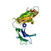
| ||||||||
|---|---|---|---|---|---|---|---|---|---|
| 1 | 
| ||||||||
| Unit cell |
| ||||||||
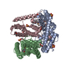
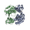
| Details | The asymmetric unit contains one chain of the Fc homodimer. The biological dimer can be generated from chain A. |
-Components
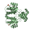
| #1: Antibody | Mass: 24821.018 Da / Num. of mol.: 1 / Fragment: CEPSILON3-CEPSILON4 Source method: isolated from a genetically manipulated source Source: (gene. exp.) Homo sapiens (human) / Plasmid: PACGP67A / Production host:  Trichoplusia ni (cabbage looper) / References: UniProt: P01854 Trichoplusia ni (cabbage looper) / References: UniProt: P01854 |
|---|---|
| #2: Water | ChemComp-HOH /  Mass: 18.015 Da / Num. of mol.: 145 / Source method: isolated from a natural source / Formula: H2O Mass: 18.015 Da / Num. of mol.: 145 / Source method: isolated from a natural source / Formula: H2O |
| Has protein modification | Y |
-Experimental details
-Experiment
| Experiment | Method: X-RAY DIFFRACTION / Number of used crystals: 1 |
|---|
- Sample preparation
| Crystal | Density Matthews: 2.64 Å3/Da / Density % sol: 45.5 % | ||||||||||||||||||||||||||||||
|---|---|---|---|---|---|---|---|---|---|---|---|---|---|---|---|---|---|---|---|---|---|---|---|---|---|---|---|---|---|---|---|
| Crystal grow | Temperature: 295 K / Method: vapor diffusion, hanging drop / pH: 4.6 Details: PEG 4000, sodium acetate pH 4.6, VAPOR DIFFUSION, HANGING DROP, temperature 295K | ||||||||||||||||||||||||||||||
| Crystal grow | *PLUS Temperature: 22 ℃ / pH: 8 | ||||||||||||||||||||||||||||||
| Components of the solutions | *PLUS
|
-Data collection
| Diffraction | Mean temperature: 113 K |
|---|---|
| Diffraction source | Source: SYNCHROTRON / Site: APS  / Beamline: 5ID-B / Wavelength: 0.906 / Beamline: 5ID-B / Wavelength: 0.906 |
| Detector | Type: MARRESEARCH / Detector: CCD / Date: Aug 7, 1999 / Details: 1mm x 1mm slits, 25 m from crystal |
| Radiation | Monochromator: Si (111), cryogenically-cooled / Protocol: SINGLE WAVELENGTH / Monochromatic (M) / Laue (L): M / Scattering type: x-ray |
| Radiation wavelength | Wavelength: 0.906 Å / Relative weight: 1 |
| Reflection | Resolution: 2.3→30 Å / Num. all: 12322 / Num. obs: 12322 / % possible obs: 99.8 % / Redundancy: 8.7 % / Biso Wilson estimate: 49.2 Å2 / Rmerge(I) obs: 0.054 / Net I/σ(I): 33.9 |
| Reflection shell | Resolution: 2.3→2.38 Å / Redundancy: 7.1 % / Rmerge(I) obs: 0.257 / Mean I/σ(I) obs: 10.6 / Num. unique all: 1208 / % possible all: 100 |
| Reflection | *PLUS Highest resolution: 2.3 Å / Num. obs: 12340 / Num. measured all: 106855 |
| Reflection shell | *PLUS % possible obs: 100 % |
- Processing
| Software |
| ||||||||||||||||||||||||||||||||||||
|---|---|---|---|---|---|---|---|---|---|---|---|---|---|---|---|---|---|---|---|---|---|---|---|---|---|---|---|---|---|---|---|---|---|---|---|---|---|
| Refinement | Method to determine structure: MOLECULAR REPLACEMENT / Resolution: 2.3→30 Å / σ(F): 0 / Stereochemistry target values: Engh & Huber Details: Density for the Cepsilon4 AB loop was particularly poor, especially from residues 453-457, and this region of the loop was built sterically. The B-factors were generally higher and the ...Details: Density for the Cepsilon4 AB loop was particularly poor, especially from residues 453-457, and this region of the loop was built sterically. The B-factors were generally higher and the electron density generally poorer for the BC- DE- and FG-loops of the Cepsilon3 domain that bind to the high affinity receptor. The residue K435 sits at the interdomain interface and the side chain may partially fill a gap in the space between the domains. This is not obvious from the structure, however, as all of the atoms could not be modeled for this residue. The structure contains two cis-Prolines : P471 and P533 There are N-linked carbohydrates attached to the protein at residues N371 and N394. Some electron density is observed for the carbohydrate attached to N394, but no electron density is observed for the carbohydrate attached to N371. Carbohydrates were not modeled in the structure and so they are not present in the PDB file.
| ||||||||||||||||||||||||||||||||||||
| Displacement parameters | Biso mean: 51.9 Å2
| ||||||||||||||||||||||||||||||||||||
| Refinement step | Cycle: LAST / Resolution: 2.3→30 Å
| ||||||||||||||||||||||||||||||||||||
| Refine LS restraints |
| ||||||||||||||||||||||||||||||||||||
| Software | *PLUS Name: CNS / Classification: refinement | ||||||||||||||||||||||||||||||||||||
| Refinement | *PLUS Lowest resolution: 30 Å / Num. reflection obs: 11053 / σ(F): 0 / % reflection Rfree: 10.3 % / Rfactor obs: 0.242 / Rfactor Rfree: 0.27 | ||||||||||||||||||||||||||||||||||||
| Solvent computation | *PLUS | ||||||||||||||||||||||||||||||||||||
| Displacement parameters | *PLUS Biso mean: 51.9 Å2 | ||||||||||||||||||||||||||||||||||||
| Refine LS restraints | *PLUS
|
