 Movie
Movie Controller

[English] 日本語
 Yorodumi
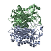
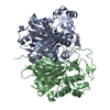
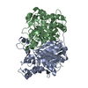
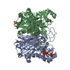
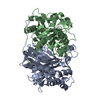
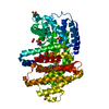
- PDB-3h76: Crystal structure of PqsD, a key enzyme in Pseudomonas aeruginosa... -
+ Open data

- Basic information
| Entry | Database: PDB / ID: 3h76 | ||||||
|---|---|---|---|---|---|---|---|
| Title | Crystal structure of PqsD, a key enzyme in Pseudomonas aeruginosa quinolone signal biosynthesis pathway | ||||||
 Components Components | PQS biosynthetic enzyme | ||||||
 Keywords Keywords | TRANSFERASE / PqsD / PQS / ANTHRANILOYL-CoA / ANTHRANILIC ACID | ||||||
| Function / homology |  Function and homology information Function and homology informationanthraniloyl-CoA anthraniloyltransferase / beta-ketoacyl-acyl-carrier-protein synthase III activity / fatty acid elongation / secondary metabolite biosynthetic process / acyltransferase activity / 3-oxoacyl-[acyl-carrier-protein] synthase activity / cytoplasm Similarity search - Function | ||||||
| Biological species |  Pseudomonas aeruginosa PAO1 (bacteria) Pseudomonas aeruginosa PAO1 (bacteria) | ||||||
| Method |  X-RAY DIFFRACTION / MOLECULAR REPLACEMENT / Resolution: 1.8 Å X-RAY DIFFRACTION / MOLECULAR REPLACEMENT / Resolution: 1.8 Å | ||||||
 Authors Authors | Bera, A.K. / Atanasova, V. / Parsons, J.F. | ||||||
 Citation Citation | Journal: Biochemistry / Year: 2009 Title: Structure of PqsD, a Pseudomonas quinolone signal biosynthetic enzyme, in complex with anthranilate. Authors: Bera, A.K. / Atanasova, V. / Robinson, H. / Eisenstein, E. / Coleman, J.P. / Pesci, E.C. / Parsons, J.F. | ||||||
| History |
|
- Structure visualization
| Structure viewer | Molecule: MolmilJmol/JSmol |
|---|
- Downloads & links
-Download
| PDBx/mmCIF format | 3h76.cif.gz | 263.5 KB | Display | PDBx/mmCIF format |
|---|---|---|---|---|
| PDB format | pdb3h76.ent.gz | 212.2 KB | Display | PDB format |
| PDBx/mmJSON format | 3h76.json.gz | Tree view | PDBx/mmJSON format | |
| Others |  Other downloads Other downloads |
-Validation report
| Arichive directory | https://data.pdbj.org/pub/pdb/validation_reports/h7/3h76ftp://data.pdbj.org/pub/pdb/validation_reports/h7/3h76 | HTTPS FTP |
|---|
-Related structure data
| Related structure data | 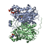 3h77C 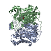 3h78C 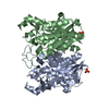 1hn9S C: citing same article ( S: Starting model for refinement |
|---|---|
| Similar structure data |


-Links
 PDBj
PDBj

- Assembly
| Deposited unit | 
| ||||||||
|---|---|---|---|---|---|---|---|---|---|
| 1 |
| ||||||||
| Unit cell |
|
-Components
| #1: Protein | Mass: 38744.227 Da / Num. of mol.: 2 Source method: isolated from a genetically manipulated source Source: (gene. exp.) Pseudomonas aeruginosa PAO1 (bacteria) / Strain: PA01 / 1C / PRS 101 / LMG 12228 / Gene: pqsD, PA0999 / Plasmid: pET28a / Production host: References: UniProt: P20582, beta-ketoacyl-[acyl-carrier-protein] synthase III #2: Water | ChemComp-HOH / |  Mass: 18.015 Da / Num. of mol.: 348 / Source method: isolated from a natural source / Formula: H2O Mass: 18.015 Da / Num. of mol.: 348 / Source method: isolated from a natural source / Formula: H2O |
|---|
-Experimental details
-Experiment
| Experiment | Method: X-RAY DIFFRACTION / Number of used crystals: 1 |
|---|
- Sample preparation
| Crystal | Density Matthews: 1.94 Å3/Da / Density % sol: 36.45 % |
|---|---|
| Crystal grow | Temperature: 293 K / Method: vapor diffusion, hanging drop / pH: 8.5 Details: 0.16 M MgCl2, 0.08M Tris-HCl pH 8.5, 24% PEG 4000, 20% Glycerol, VAPOR DIFFUSION, HANGING DROP, temperature 293K |
-Data collection
| Diffraction | Mean temperature: 100 K |
|---|---|
| Diffraction source | Source: ROTATING ANODE / Type: RIGAKU MICROMAX-007 / Wavelength: 1.5418 Å |
| Detector | Type: MAR scanner 345 mm plate / Detector: IMAGE PLATE / Date: Sep 18, 2008 |
| Radiation | Protocol: SINGLE WAVELENGTH / Monochromatic (M) / Laue (L): M / Scattering type: x-ray |
| Radiation wavelength | Wavelength: 1.5418 Å / Relative weight: 1 |
| Reflection | Resolution: 1.8→27 Å / Num. obs: 49952 / % possible obs: 91.4 % / Observed criterion σ(F): 0 / Observed criterion σ(I): 0 / Redundancy: 3.1 % / Rmerge(I) obs: 0.091 / Net I/σ(I): 5.1 |
| Reflection shell | Resolution: 1.8→1.86 Å / Redundancy: 3.07 % / Rmerge(I) obs: 0.51 / Mean I/σ(I) obs: 1.6 / % possible all: 88.3 |
- Processing
| Software |
| |||||||||||||||||||||||||||||||||||||||||||||||||||||||||||||||||||||||||||||||||||||||||||||||||||||||||
|---|---|---|---|---|---|---|---|---|---|---|---|---|---|---|---|---|---|---|---|---|---|---|---|---|---|---|---|---|---|---|---|---|---|---|---|---|---|---|---|---|---|---|---|---|---|---|---|---|---|---|---|---|---|---|---|---|---|---|---|---|---|---|---|---|---|---|---|---|---|---|---|---|---|---|---|---|---|---|---|---|---|---|---|---|---|---|---|---|---|---|---|---|---|---|---|---|---|---|---|---|---|---|---|---|---|---|
| Refinement | Method to determine structure: MOLECULAR REPLACEMENT Starting model: PDB entry 1HN9 Resolution: 1.8→25.6 Å / Cor.coef. Fo:Fc: 0.967 / Cor.coef. Fo:Fc free: 0.942 / SU B: 11.617 / SU ML: 0.151 / Cross valid method: THROUGHOUT / ESU R Free: 0.17 / Stereochemistry target values: MAXIMUM LIKELIHOOD / Details: HYDROGENS HAVE BEEN ADDED IN THE RIDING POSITIONS
| |||||||||||||||||||||||||||||||||||||||||||||||||||||||||||||||||||||||||||||||||||||||||||||||||||||||||
| Solvent computation | Ion probe radii: 0.8 Å / Shrinkage radii: 0.8 Å / VDW probe radii: 1.2 Å / Solvent model: MASK | |||||||||||||||||||||||||||||||||||||||||||||||||||||||||||||||||||||||||||||||||||||||||||||||||||||||||
| Displacement parameters | Biso mean: 34.725 Å2
| |||||||||||||||||||||||||||||||||||||||||||||||||||||||||||||||||||||||||||||||||||||||||||||||||||||||||
| Refinement step | Cycle: LAST / Resolution: 1.8→25.6 Å
| |||||||||||||||||||||||||||||||||||||||||||||||||||||||||||||||||||||||||||||||||||||||||||||||||||||||||
| Refine LS restraints |
| |||||||||||||||||||||||||||||||||||||||||||||||||||||||||||||||||||||||||||||||||||||||||||||||||||||||||
| LS refinement shell | Resolution: 1.8→1.847 Å / Total num. of bins used: 20
|
