Movie
Movie Controller

[English] 日本語
 Yorodumi
- EMDB-4939: Structure of PTCH1 bound to a modified Hedgehog ligand ShhN-C24II... -
+ Open data

- Basic information
| Entry | Database: EMDB / ID: EMD-4939 | |||||||||
|---|---|---|---|---|---|---|---|---|---|---|
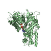
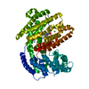
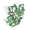
| Title | Structure of PTCH1 bound to a modified Hedgehog ligand ShhN-C24II: focused refinement of the membrane domain | |||||||||
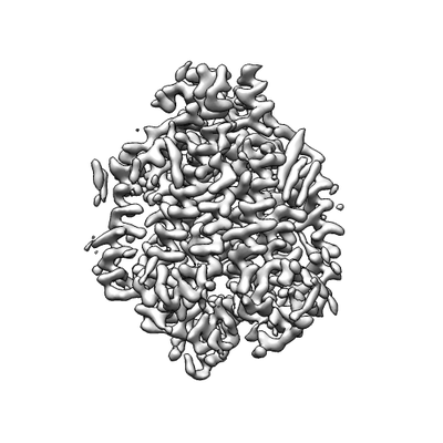
 Map data Map data | Focused refinement of the membrane domain of PTCH1 | |||||||||
 Sample Sample |
| |||||||||
| Function / homology |  Function and homology information Function and homology informationneural plate axis specification / response to chlorate / cell differentiation involved in kidney development / cell proliferation involved in metanephros development / hedgehog receptor activity / neural tube patterning / smoothened binding / hedgehog family protein binding / hindlimb morphogenesis / Ligand-receptor interactions ...neural plate axis specification / response to chlorate / cell differentiation involved in kidney development / cell proliferation involved in metanephros development / hedgehog receptor activity / neural tube patterning / smoothened binding / hedgehog family protein binding / hindlimb morphogenesis / Ligand-receptor interactions / epidermal cell fate specification / spinal cord motor neuron differentiation / prostate gland development / negative regulation of cell division / patched binding / somite development / limb morphogenesis / Activation of SMO / smooth muscle tissue development / pharyngeal system development / mammary gland duct morphogenesis / mammary gland epithelial cell differentiation / cellular response to cholesterol / cell fate determination / metanephric collecting duct development / commissural neuron axon guidance / response to alkaloid / regulation of smoothened signaling pathway / dorsal/ventral pattern formation / Class B/2 (Secretin family receptors) / embryonic limb morphogenesis / negative regulation of multicellular organism growth / branching involved in ureteric bud morphogenesis / cholesterol binding / dorsal/ventral neural tube patterning / ciliary membrane / positive regulation of epidermal cell differentiation / dendritic growth cone / keratinocyte proliferation / positive regulation of cholesterol efflux / negative regulation of keratinocyte proliferation / spermatid development / response to retinoic acid / embryonic organ development / response to mechanical stimulus / negative regulation of osteoblast differentiation / negative regulation of stem cell proliferation / axonal growth cone / heart morphogenesis / Hedgehog 'off' state / regulation of mitotic cell cycle / liver regeneration / stem cell proliferation / cyclin binding / animal organ morphogenesis / protein localization to plasma membrane / negative regulation of smoothened signaling pathway / neural tube closure / protein processing / brain development / caveola / Hedgehog 'on' state / apical part of cell / endocytic vesicle membrane / response to estradiol / heparin binding / regulation of protein localization / glucose homeostasis / midbody / in utero embryonic development / postsynaptic membrane / response to xenobiotic stimulus / positive regulation of DNA-templated transcription / protein-containing complex binding / perinuclear region of cytoplasm / negative regulation of transcription by RNA polymerase II / signal transduction / plasma membrane Similarity search - Function | |||||||||
| Biological species |  Homo sapiens (human) Homo sapiens (human) | |||||||||
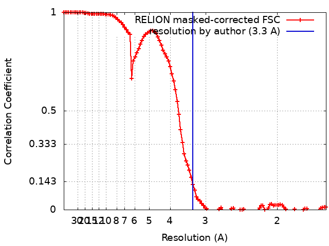
| Method | single particle reconstruction / cryo EM / Resolution: 3.3 Å | |||||||||
 Authors Authors | Korkhov VM / Qi C | |||||||||
| Funding support |  Switzerland, 1 items Switzerland, 1 items
| |||||||||
 Citation Citation | Journal: Sci Adv / Year: 2019 Title: Structural basis of sterol recognition by human hedgehog receptor PTCH1. Authors: Chao Qi / Giulio Di Minin / Irene Vercellino / Anton Wutz / Volodymyr M Korkhov / Abstract: Hedgehog signaling is central in embryonic development and tissue regeneration. Disruption of the pathway is linked to genetic diseases and cancer. Binding of the secreted ligand, Sonic hedgehog ...Hedgehog signaling is central in embryonic development and tissue regeneration. Disruption of the pathway is linked to genetic diseases and cancer. Binding of the secreted ligand, Sonic hedgehog (ShhN) to its receptor Patched (PTCH1) activates the signaling pathway. Here, we describe a 3.4-Å cryo-EM structure of the human PTCH1 bound to ShhN, a modified hedgehog ligand mimicking its palmitoylated form. The membrane-embedded part of PTCH1 is surrounded by 10 sterol molecules at the inner and outer lipid bilayer portion of the protein. The annular sterols interact at multiple sites with both the sterol-sensing domain (SSD) and the SSD-like domain (SSDL), which are located on opposite sides of PTCH1. The structure reveals a possible route for sterol translocation across the lipid bilayer by PTCH1 and homologous transporters. | |||||||||
| History |
|
- Structure visualization
| Movie |
Movie viewer |
|---|---|
| Structure viewer | EM map: SurfViewMolmilJmol/JSmol |
| Supplemental images |
- Downloads & links
-EMDB archive
| Map data | emd_4939.map.gz | 3.6 MB | EMDB map data format | |
|---|---|---|---|---|
| Header (meta data) | emd-4939-v30.xmlemd-4939.xml | 14.7 KB 14.7 KB | Display Display | EMDB header |
| FSC (resolution estimation) | emd_4939_fsc.xml | 10.7 KB | Display | FSC data file |
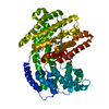
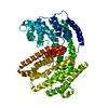
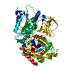
| Images |  emd_4939.png emd_4939.png | 45.4 KB | ||
| Archive directory |  http://ftp.pdbj.org/pub/emdb/structures/EMD-4939ftp://ftp.pdbj.org/pub/emdb/structures/EMD-4939 http://ftp.pdbj.org/pub/emdb/structures/EMD-4939ftp://ftp.pdbj.org/pub/emdb/structures/EMD-4939 | HTTPS FTP |
-Related structure data


-Links
| EMDB pages | EMDB (EBI/PDBe) / EMDataResource |
|---|---|
| Related items in Molecule of the Month |


-Map
| File | Download / File: emd_4939.map.gz / Format: CCP4 / Size: 103 MB / Type: IMAGE STORED AS FLOATING POINT NUMBER (4 BYTES) | ||||||||||||||||||||||||||||||||||||||||||||||||||||||||||||||||||||
|---|---|---|---|---|---|---|---|---|---|---|---|---|---|---|---|---|---|---|---|---|---|---|---|---|---|---|---|---|---|---|---|---|---|---|---|---|---|---|---|---|---|---|---|---|---|---|---|---|---|---|---|---|---|---|---|---|---|---|---|---|---|---|---|---|---|---|---|---|---|
| Annotation | Focused refinement of the membrane domain of PTCH1 | ||||||||||||||||||||||||||||||||||||||||||||||||||||||||||||||||||||
| Projections & slices | Image control
Images are generated by Spider. | ||||||||||||||||||||||||||||||||||||||||||||||||||||||||||||||||||||
| Voxel size | X=Y=Z: 0.8141 Å | ||||||||||||||||||||||||||||||||||||||||||||||||||||||||||||||||||||
| Density |
| ||||||||||||||||||||||||||||||||||||||||||||||||||||||||||||||||||||
| Symmetry | Space group: 1 | ||||||||||||||||||||||||||||||||||||||||||||||||||||||||||||||||||||
| Details | EMDB XML:
CCP4 map header:
| ||||||||||||||||||||||||||||||||||||||||||||||||||||||||||||||||||||
 Z (Sec.)
 Y (Row.)
 X (Col.)


-Supplemental data
- Sample components
-Entire : Complex of PTCH1 with a modified Hedgehog ligand ShhN-C24II
| Entire | Name: Complex of PTCH1 with a modified Hedgehog ligand ShhN-C24II |
|---|---|
| Components |
|
-Supramolecule #1: Complex of PTCH1 with a modified Hedgehog ligand ShhN-C24II
| Supramolecule | Name: Complex of PTCH1 with a modified Hedgehog ligand ShhN-C24II type: complex / ID: 1 / Parent: 0 / Macromolecule list: all |
|---|
-Supramolecule #2: Complex of PTCH1 with a modified Hedgehog ligand ShhN-C24II
| Supramolecule | Name: Complex of PTCH1 with a modified Hedgehog ligand ShhN-C24II type: complex / ID: 2 / Parent: 1 / Macromolecule list: #1 |
|---|---|
| Source (natural) | Organism: Homo sapiens (human) |
| Recombinant expression | Organism: Homo sapiens (human) |
-Supramolecule #3: Complex of PTCH1 with a modified Hedgehog ligand ShhN-C24II
| Supramolecule | Name: Complex of PTCH1 with a modified Hedgehog ligand ShhN-C24II type: complex / ID: 3 / Parent: 1 / Macromolecule list: #2 |
|---|---|
| Source (natural) | Organism: Homo sapiens (human) |
| Recombinant expression | Organism:  |
-Macromolecule #1: Protein patched homolog 1
| Macromolecule | Name: Protein patched homolog 1 / type: protein_or_peptide / ID: 1 / Enantiomer: LEVO |
|---|---|
| Source (natural) | Organism: Homo sapiens (human) |
| Recombinant expression | Organism: Homo sapiens (human) |
| Sequence | String: MASAGNAAEP QDRGGGGSGC IGAPGRPAGG GRRRRTGGLR RAAAPDRDYL HRPSYCDAAF ALEQISKGKA TGRKAPLWLR AKFQRLLFKL GCYIQKNCGK FLVVGLLIFG AFAVGLKAAN LETNVEELWV EVGGRVSREL NYTRQKIGEE AMFNPQLMIQ TPKEEGANVL ...String: MASAGNAAEP QDRGGGGSGC IGAPGRPAGG GRRRRTGGLR RAAAPDRDYL HRPSYCDAAF ALEQISKGKA TGRKAPLWLR AKFQRLLFKL GCYIQKNCGK FLVVGLLIFG AFAVGLKAAN LETNVEELWV EVGGRVSREL NYTRQKIGEE AMFNPQLMIQ TPKEEGANVL TTEALLQHLD SALQASRVHV YMYNRQWKLE HLCYKSGELI TETGYMDQII EYLYPCLIIT PLDCFWEGAK LQSGTAYLLG KPPLRWTNFD PLEFLEELKK INYQVDSWEE MLNKAEVGHG YMDRPCLNPA DPDCPATAPN KNSTKPLDMA LVLNGGCHGL SRKYMHWQEE LIVGGTVKNS TGKLVSAHAL QTMFQLMTPK QMYEHFKGYE YVSHINWNED KAAAILEAWQ RTYVEVVHQS VAQNSTQKVL SFTTTTLDDI LKSFSDVSVI RVASGYLLML AYACLTMLRW DCSKSQGAVG LAGVLLVALS VAAGLGLCSL IGISFNAATT QVLPFLALGV GVDDVFLLAH AFSETGQNKR IPFEDRTGEC LKRTGASVAL TSISNVTAFF MAALIPIPAL RAFSLQAAVV VVFNFAMVLL IFPAILSMDL YRREDRRLDI FCCFTSPCVS RVIQVEPQAY TDTHDNTRYS PPPPASSHSF AHETQITMQS TVQLRTEYDP HTHVYYTTAE PRSEISVQPV TVTQDTLSCQ SPESTSSTRD LLSQFSDSSL HCLEPPCTKW TLSSFAEKHY APFLLKPKAK VVVIFLFLGL LGVSLYGTTR VRDGLDLTDI VPRETREYDF IAAQFKYFSF YNMYIVTQKA DYPNIQHLLY DLHRSFSNVK YVMLEENKQL PKMWLHYFRD WLQGLQDAFD SDWETGKIMP NNYKNGSDDG VLAYKLLVQT GSRDKPIDIS QLTKQRLVDA DGIINPSAFY IYLTAWVSND PVAYAASQAN IRPHRPEWVH DKADYMPETR LRIPAAEPIE YAQFPFYLNG LRDTSDFVEA IEKVRTICSN YTSLGLSSYP NGYPFLFWEQ YIGLRHWLLL FISVVLACTF LVCAVFLLNP WTAGIIVMVL ALMTVELFGM MGLIGIKLSA VPVVILIASV GIGVEFTVHV ALAFLTAIGD KNRRAVLALE HMFAPVLDGA VSTLLGVLML AGSEFDFIVR YFFAVLAILT ILGVLNGLVL LPVLLSFFGP YPEVSPANAA ALEVLFQGPG GVSKGEELFT GVVPILVELD GDVNGHKFSV SGEGEGDATY GKLTLKFICT TGKLPVPWPT LVTTFGYGLQ CFARYPDHMK QHDFFKSAMP EGYVQERTIF FKDDGNYKTR AEVKFEGDTL VNRIELKGID FKEDGNILGH KLEYNYNSHN VYIMADKQKN GIKVNFKIRH NIEDGSVQLA DHYQQNTPIG DGPVLLPDNH YLSYQSALSK DPNEKRDHMV LLEFVTAAGI TLGMDELYKA ASAWSHPQFE KGGGSGGGSG GSAWSHPQFE K |
-Macromolecule #2: Sonic hedgehog protein
| Macromolecule | Name: Sonic hedgehog protein / type: protein_or_peptide / ID: 2 / Details: ShhN-C24II, a modified sonic hedgehog protein / Enantiomer: LEVO |
|---|---|
| Source (natural) | Organism: Homo sapiens (human) |
| Recombinant expression | Organism: |
| Sequence | String: MKKHHHHHHG SGMSDSEVNQ EAKPEVKPEV KPETHINLKV SDGSSEIFFK IKKTTPLRRL MEAFAKRQGK EMDSLRFLYD GIRIQADQTP EDLDMEDNDI IEAHREQIGG IIGPGRGFGK RRHPKKLTPL AYKQFIPNVA EKTLGASGRY EGKISRNSER FKELTPNYNP ...String: MKKHHHHHHG SGMSDSEVNQ EAKPEVKPEV KPETHINLKV SDGSSEIFFK IKKTTPLRRL MEAFAKRQGK EMDSLRFLYD GIRIQADQTP EDLDMEDNDI IEAHREQIGG IIGPGRGFGK RRHPKKLTPL AYKQFIPNVA EKTLGASGRY EGKISRNSER FKELTPNYNP DIIFKDEENT GADRLMTQRC KDKLNALAIS VMNQWPGVKL RVTEGWDEDG HHSEESLHYE GRAVDITTSD RDRSKYGMLA RLAVEAGFDW VYYESKAHIH CSVKAENSVA AKSGG |
-Experimental details
-Structure determination
| Method | cryo EM |
|---|---|
 Processing Processing | single particle reconstruction |
| Aggregation state | particle |
-Sample preparation
| Buffer | pH: 8 |
|---|---|
| Vitrification | Cryogen name: ETHANE / Instrument: FEI VITROBOT MARK IV |
- Electron microscopy
| Microscope | FEI TITAN KRIOS |
|---|---|
| Image recording | Film or detector model: GATAN K2 QUANTUM (4k x 4k) / Detector mode: COUNTING / Average electron dose: 44.7 e/Å2 |
| Electron beam | Acceleration voltage: 300 kV / Electron source:  FIELD EMISSION GUN FIELD EMISSION GUN |
| Electron optics | Illumination mode: FLOOD BEAM / Imaging mode: BRIGHT FIELD / Nominal defocus max: -2.4 µm / Nominal defocus min: -0.8 µm |
| Sample stage | Specimen holder model: FEI TITAN KRIOS AUTOGRID HOLDER / Cooling holder cryogen: NITROGEN |
| Experimental equipment |  Model: Titan Krios / Image courtesy: FEI Company |