 Movie
Movie Controller


+ Open data

- Basic information
| Entry | Database: PDB / ID: 3h34 | ||||||
|---|---|---|---|---|---|---|---|
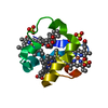
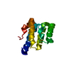
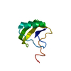
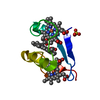
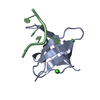
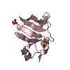
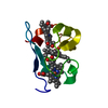
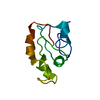
| Title | PpcE, A cytochrome c7 from Geobacter sulfurreducens | ||||||
 Components Components | Cytochrome c7 | ||||||
 Keywords Keywords | ELECTRON TRANSPORT / cytochrome c7 / Multiheme cytochrome / Geobacter sulfurreducens | ||||||
| Function / homology |  Function and homology information Function and homology information | ||||||
| Biological species |  Geobacter sulfurreducens (bacteria) Geobacter sulfurreducens (bacteria) | ||||||
| Method |  X-RAY DIFFRACTION / SYNCHROTRON / MAD / Resolution: 1.6 Å X-RAY DIFFRACTION / SYNCHROTRON / MAD / Resolution: 1.6 Å | ||||||
 Authors Authors | Pokkuluri, P.R. / Schiffer, M. | ||||||
 Citation Citation | Journal: Biochim.Biophys.Acta / Year: 2010 Title: Structural characterization of a family of cytochromes c(7) involved in Fe(III) respiration by Geobacter sulfurreducens. Authors: Pokkuluri, P.R. / Londer, Y.Y. / Yang, X. / Duke, N.E. / Erickson, J. / Orshonsky, V. / Johnson, G. / Schiffer, M. | ||||||
| History |
|
- Structure visualization
| Structure viewer | Molecule: MolmilJmol/JSmol |
|---|
- Downloads & links
-Download
| PDBx/mmCIF format | 3h34.cif.gz | 32.9 KB | Display | PDBx/mmCIF format |
|---|---|---|---|---|
| PDB format | pdb3h34.ent.gz | 21.7 KB | Display | PDB format |
| PDBx/mmJSON format | 3h34.json.gz | Tree view | PDBx/mmJSON format | |
| Others |  Other downloads Other downloads |
-Validation report
| Arichive directory | https://data.pdbj.org/pub/pdb/validation_reports/h3/3h34ftp://data.pdbj.org/pub/pdb/validation_reports/h3/3h34 | HTTPS FTP |
|---|
-Related structure data


-Links
 PDBj
PDBj
- Assembly
| Deposited unit | 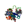
| ||||||||
|---|---|---|---|---|---|---|---|---|---|
| 1 |
| ||||||||
| Unit cell |
| ||||||||
| Details | Monomer per asymmetric unit; Monomer in solution based on size exclusion column chromatography and dynamic light scattering |
-Components
| #1: Protein | Mass: 7833.074 Da / Num. of mol.: 1 Source method: isolated from a genetically manipulated source Source: (gene. exp.) Geobacter sulfurreducens (bacteria) / Gene: cyd-5, GSU1760 / Production host: | ||
|---|---|---|---|
| #2: Chemical |   Mass: 616.487 Da / Num. of mol.: 3 / Source method: obtained synthetically / Formula: C34H32FeN4O4 Mass: 616.487 Da / Num. of mol.: 3 / Source method: obtained synthetically / Formula: C34H32FeN4O4#3: Water | ChemComp-HOH / |  Mass: 18.015 Da / Num. of mol.: 103 / Source method: isolated from a natural source / Formula: H2O Mass: 18.015 Da / Num. of mol.: 103 / Source method: isolated from a natural source / Formula: H2O |
-Experimental details
-Experiment
| Experiment | Method: X-RAY DIFFRACTION / Number of used crystals: 1 |
|---|
- Sample preparation
| Crystal | Density Matthews: 2.79 Å3/Da / Density % sol: 55.85 % |
|---|---|
| Crystal grow | Temperature: 298 K / Method: vapor diffusion, hanging drop / pH: 7.8 Details: Micro crystals obtained from SaltRx-31 (3.5 M sodium formate, 0.1 M bis-tris propane, pH 7.0) were optimized by micro seeding method, VAPOR DIFFUSION, HANGING DROP, temperature 298K |
-Data collection
| Diffraction | Mean temperature: 100 K | |||||||||||||||
|---|---|---|---|---|---|---|---|---|---|---|---|---|---|---|---|---|
| Diffraction source | Source: SYNCHROTRON / Site: APS  / Beamline: 19-BM / Wavelength: 1.73859, 1.73647, 1.78875, 1.0332 / Beamline: 19-BM / Wavelength: 1.73859, 1.73647, 1.78875, 1.0332 | |||||||||||||||
| Detector | Type: CUSTOM-MADE / Detector: CCD / Date: Aug 25, 2003 | |||||||||||||||
| Radiation | Protocol: MAD / Monochromatic (M) / Laue (L): M / Scattering type: x-ray | |||||||||||||||
| Radiation wavelength |
| |||||||||||||||
| Reflection | Resolution: 1.6→50 Å / Num. all: 22388 / Num. obs: 22388 / % possible obs: 99 % / Redundancy: 4 % / Biso Wilson estimate: 11.5 Å2 / Rmerge(I) obs: 0.079 / Net I/σ(I): 28.9 | |||||||||||||||
| Reflection shell | Resolution: 1.6→1.66 Å / Redundancy: 3 % / Rmerge(I) obs: 0.313 / Mean I/σ(I) obs: 5.5 / Num. unique all: 2226 / % possible all: 98 |
- Processing
| Software |
| ||||||||||||||||||||||||||||||||||||||||||||||||||||||||||||||||||||||||||||||||
|---|---|---|---|---|---|---|---|---|---|---|---|---|---|---|---|---|---|---|---|---|---|---|---|---|---|---|---|---|---|---|---|---|---|---|---|---|---|---|---|---|---|---|---|---|---|---|---|---|---|---|---|---|---|---|---|---|---|---|---|---|---|---|---|---|---|---|---|---|---|---|---|---|---|---|---|---|---|---|---|---|---|
| Refinement | Method to determine structure: MAD / Resolution: 1.6→30.32 Å / Rfactor Rfree error: 0.005 / Data cutoff high absF: 624877.33 / Data cutoff low absF: 0 / Isotropic thermal model: RESTRAINED / Cross valid method: THROUGHOUT / σ(F): 0 / Stereochemistry target values: Engh & Huber
| ||||||||||||||||||||||||||||||||||||||||||||||||||||||||||||||||||||||||||||||||
| Solvent computation | Solvent model: FLAT MODEL / Bsol: 74.2616 Å2 / ksol: 0.445928 e/Å3 | ||||||||||||||||||||||||||||||||||||||||||||||||||||||||||||||||||||||||||||||||
| Displacement parameters | Biso mean: 13.3 Å2
| ||||||||||||||||||||||||||||||||||||||||||||||||||||||||||||||||||||||||||||||||
| Refine analyze |
| ||||||||||||||||||||||||||||||||||||||||||||||||||||||||||||||||||||||||||||||||
| Refinement step | Cycle: LAST / Resolution: 1.6→30.32 Å
| ||||||||||||||||||||||||||||||||||||||||||||||||||||||||||||||||||||||||||||||||
| Refine LS restraints |
| ||||||||||||||||||||||||||||||||||||||||||||||||||||||||||||||||||||||||||||||||
| LS refinement shell | Resolution: 1.6→1.7 Å / Rfactor Rfree error: 0.015 / Total num. of bins used: 6
| ||||||||||||||||||||||||||||||||||||||||||||||||||||||||||||||||||||||||||||||||
| Xplor file |
|
