| 登録情報 | データベース: PDB / ID: 3h1d
|
|---|
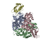
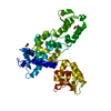
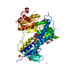
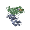
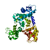
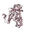
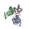
| タイトル | Structure of the HUWE1 HECT Domain |
|---|
 要素 要素 | E3 ubiquitin-protein ligase HUWE1 |
|---|
 キーワード キーワード | LIGASE / E3Ligase / ubiquitin / HECT / lobe / Alternative splicing / Chromosomal rearrangement / Cytoplasm / Differentiation / Disease mutation / DNA-binding / Mental retardation / Nucleus / Phosphoprotein / Ubl conjugation pathway |
|---|
| 機能・相同性 |  機能・相同性情報 機能・相同性情報
negative regulation of peroxisome proliferator activated receptor signaling pathway / histone ubiquitin ligase activity / negative regulation of mitochondrial fusion / protein branched polyubiquitination / positive regulation of type 2 mitophagy / HECT-type E3 ubiquitin transferase / : / ubiquitin-ubiquitin ligase activity / Golgi organization / protein monoubiquitination ...negative regulation of peroxisome proliferator activated receptor signaling pathway / histone ubiquitin ligase activity / negative regulation of mitochondrial fusion / protein branched polyubiquitination / positive regulation of type 2 mitophagy / HECT-type E3 ubiquitin transferase / : / ubiquitin-ubiquitin ligase activity / Golgi organization / protein monoubiquitination / protein K48-linked ubiquitination / positive regulation of protein ubiquitination / circadian regulation of gene expression / base-excision repair / protein polyubiquitination / ubiquitin-protein transferase activity / ubiquitin protein ligase activity / Antigen processing: Ubiquitination & Proteasome degradation / nuclear membrane / secretory granule lumen / ficolin-1-rich granule lumen / ubiquitin-dependent protein catabolic process / membrane fusion / proteasome-mediated ubiquitin-dependent protein catabolic process / positive regulation of canonical NF-kappaB signal transduction / cell differentiation / Golgi membrane / Neutrophil degranulation / mitochondrion / DNA binding / RNA binding / extracellular exosome / extracellular region / nucleoplasm / membrane / nucleus / cytosol / cytoplasm類似検索 - 分子機能 Hect, E3 ligase catalytic domains / Hect, E3 ligase catalytic domain fold / Hect, E3 ligase catalytic domain / Hect, E3 ligase catalytic domain / HUWE1, UBA domain / Hect, E3 ligase catalytic fold / Hect, E3 ligase catalytic domain / E3 ubiquitin ligase, domain of unknown function DUF908 / E3 ubiquitin ligase, domain of unknown function DUF913 / Domain of Unknown Function (DUF908) ...Hect, E3 ligase catalytic domains / Hect, E3 ligase catalytic domain fold / Hect, E3 ligase catalytic domain / Hect, E3 ligase catalytic domain / HUWE1, UBA domain / Hect, E3 ligase catalytic fold / Hect, E3 ligase catalytic domain / E3 ubiquitin ligase, domain of unknown function DUF908 / E3 ubiquitin ligase, domain of unknown function DUF913 / Domain of Unknown Function (DUF908) / Domain of Unknown Function (DUF913) / WWE domain / UBA-like domain / WWE domain superfamily / WWE domain / WWE domain profile. / HUWE1/Rev1, ubiquitin binding region / Ubiquitin binding region / Ubiquitin-binding motif (UBM) domain profile. / : / HECT domain / HECT, E3 ligase catalytic domain / HECT-domain (ubiquitin-transferase) / HECT domain profile. / Domain Homologous to E6-AP Carboxyl Terminus with / Ubiquitin associated domain / Ubiquitin-associated domain / Ubiquitin-associated domain (UBA) profile. / UBA-like superfamily / Armadillo-type fold / Alpha-Beta Complex / 2-Layer Sandwich / Alpha Beta類似検索 - ドメイン・相同性 |
|---|
| 生物種 |  Homo sapiens (ヒト) Homo sapiens (ヒト) |
|---|
| 手法 |  X線回折 / シンクロトロン / 分子置換 / 解像度: 1.892 Å X線回折 / シンクロトロン / 分子置換 / 解像度: 1.892 Å |
|---|
 データ登録者 データ登録者 | Partridge, J.R. / Schwartz, T.U. |
|---|
 引用 引用 | ジャーナル: J.Biol.Chem. / 年: 2010
タイトル: A structural element within the HUWE1 HECT domain modulates self-ubiquitination and substrate ubiquitination activities.
著者: Pandya, R.K. / Partridge, J.R. / Love, K.R. / Schwartz, T.U. / Ploegh, H.L. |
|---|
| 履歴 | | 登録 | 2009年4月11日 | 登録サイト: RCSB / 処理サイト: RCSB |
|---|
| 改定 1.0 | 2009年12月8日 | Provider: repository / タイプ: Initial release |
|---|
| 改定 1.1 | 2011年7月13日 | Group: Version format compliance |
|---|
| 改定 1.2 | 2021年10月13日 | Group: Database references / Derived calculations / カテゴリ: database_2 / struct_ref_seq_dif / struct_site
Item: _database_2.pdbx_DOI / _database_2.pdbx_database_accession ..._database_2.pdbx_DOI / _database_2.pdbx_database_accession / _struct_ref_seq_dif.details / _struct_site.pdbx_auth_asym_id / _struct_site.pdbx_auth_comp_id / _struct_site.pdbx_auth_seq_id |
|---|
| 改定 1.3 | 2023年9月6日 | Group: Data collection / Refinement description
カテゴリ: chem_comp_atom / chem_comp_bond / pdbx_initial_refinement_model |
|---|
|
|---|
 ムービー
ムービー コントローラー


 データを開く
 基本情報
基本情報 構造の表示
構造の表示 ダウンロードとリンク
 その他のダウンロード


 PDBj
PDBj

 集合体
集合体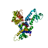


 分子量: 96.063 Da / 分子数: 4 / 由来タイプ: 合成 / 式: SO4
 分子量: 18.015 Da / 分子数: 357 / 由来タイプ: 天然 / 式: H2O
 試料調製
試料調製 / ビームライン: 24-ID-C / 波長: 0.979 Å
 解析
解析