[English] 日本語
Yorodumi
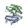
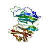
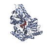
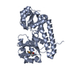
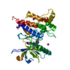
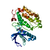
- PDB-3gyd: Crystal structure of a cyclic nucleotide-binding domain (mfla_192... -

+
Open data


ID or keywords:

Loading...

-
Basic information

Entry
Database: PDB / ID: 3gyd
TitleCrystal structure of a cyclic nucleotide-binding domain (mfla_1926) from methylobacillus flagellatus kt at 1.79 A resolution
ComponentsCyclic nucleotide-binding domain
KeywordsDNA BINDING PROTEIN / Nucleotide binding protein / structural genomics / Joint Center for Structural Genomics / JCSG / Protein Structure Initiative / PSI-2
Function / homology
Function and homology information


Cyclic nucleotide-monophosphate binding domain / Cyclic nucleotide-binding domain / cAMP/cGMP binding motif profile. / Cyclic nucleotide-binding domain / Cyclic nucleotide-binding domain superfamily / Jelly Rolls / RmlC-like jelly roll fold / Jelly Rolls / Sandwich / Mainly Beta
Similarity search - Domain/homology
ADENOSINE-3',5'-CYCLIC-MONOPHOSPHATE / Cyclic nucleotide-binding domain (cNMP-BD) protein
Similarity search - Component
Biological speciesMethylobacillus flagellatus KT (bacteria)
MethodX-RAY DIFFRACTION / SYNCHROTRON / MAD / Resolution: 1.79 Å
AuthorsJoint Center for Structural Genomics (JCSG)
CitationJournal: To be published
Title: Crystal structure of cyclic nucleotide-binding domain (YP_546034.1) from Methylobacillus flagellatus KT at 1.79 A resolution
Authors: Joint Center for Structural Genomics (JCSG)
History
DepositionApr 3, 2009Deposition site: RCSB / Processing site: RCSB
Revision 1.0Apr 21, 2009Provider: repository / Type: Initial release
Revision 1.1Jul 13, 2011Group: Advisory / Version format compliance
Revision 1.2Nov 1, 2017Group: Refinement description / Category: software / Item: _software.classification / _software.name
Revision 1.3Jul 24, 2019Group: Data collection / Derived calculations / Refinement description
Category: software / struct_conn
Item: _software.classification / _software.contact_author ..._software.classification / _software.contact_author / _software.contact_author_email / _software.language / _software.location / _software.name / _software.type / _software.version / _struct_conn.pdbx_leaving_atom_flag
Revision 1.4Feb 1, 2023Group: Database references / Derived calculations / Category: database_2 / struct_ref_seq_dif / struct_site
Item: _database_2.pdbx_DOI / _database_2.pdbx_database_accession ..._database_2.pdbx_DOI / _database_2.pdbx_database_accession / _struct_ref_seq_dif.details / _struct_site.pdbx_auth_asym_id / _struct_site.pdbx_auth_comp_id / _struct_site.pdbx_auth_seq_id

-
Structure visualization

Structure viewerMolecule:
MolmilJmol/JSmol

Downloads & links

-
Assembly

Deposited unit
A: Cyclic nucleotide-binding domain
B: Cyclic nucleotide-binding domain
hetero molecules


Theoretical massNumber of molelcules
Total (without water)43,3945
Polymers42,6432
Non-polymers7513
Water4,612256
1


  • Idetical with deposited unit
  • defined by software
TypeNameSymmetry operationNumber
identity operation1_555x,y,z1
Buried area5180 Å2
ΔGint-39.4 kcal/mol
Surface area14810 Å2
MethodPISA
Unit cell
Length a, b, c (Å)60.032, 39.745, 70.977
Angle α, β, γ (deg.)90.000, 101.850, 90.000
Int Tables number4
Space group name H-MP1211
Noncrystallographic symmetry (NCS)NCS domain:
IDEns-IDDetails
11A
21B

NCS domain segments:
Dom-IDComponent-IDEns-IDRefine codeAuth asym-IDAuth seq-ID
1116A5 - 164
2116B5 - 164

-
Components

#1: Protein Cyclic nucleotide-binding domain / / CNMP-BD protein


Mass: 21321.631 Da / Num. of mol.: 2
Source method: isolated from a genetically manipulated source
Source: (gene. exp.) Methylobacillus flagellatus KT (bacteria)
Strain: KT / DSM 6875 / Gene: Mfla_1926, YP_546034.1 / Plasmid: SpeedET / Production host: Escherichia coli (E. coli) / Strain (production host): HK100 / References: UniProt: Q1GZZ4
#2: Chemical ChemComp-CMP / ADENOSINE-3',5'-CYCLIC-MONOPHOSPHATE / CYCLIC AMP / CAMP / Cyclic adenosine monophosphate


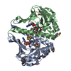
Mass: 329.206 Da / Num. of mol.: 2 / Source method: obtained synthetically / Formula: C10H12N5O6P
#3: Chemical ChemComp-GOL / GLYCEROL / GLYCERIN / PROPANE-1,2,3-TRIOL / Glycerol


Mass: 92.094 Da / Num. of mol.: 1 / Source method: obtained synthetically / Formula: C3H8O3
#4: Water ChemComp-HOH / water / Water


Mass: 18.015 Da / Num. of mol.: 256 / Source method: isolated from a natural source / Formula: H2O
Sequence detailsTHE CONSTRUCT WAS EXPRESSED WITH A PURIFICATION TAG MGSDKIHHHHHHENLYFQG.

-
Experimental details

-
Experiment

ExperimentMethod: X-RAY DIFFRACTION / Number of used crystals: 1

-
Sample preparation

CrystalDensity Matthews: 1.94 Å3/Da / Density % sol: 36.71 %
Crystal growTemperature: 277 K / Method: vapor diffusion, sitting drop / pH: 10
Details: NANODROP, 36.0% PEG 3000, 0.1M CHES pH 10.0, VAPOR DIFFUSION, SITTING DROP, temperature 277K

-
Data collection

DiffractionMean temperature: 100 K
Diffraction sourceSource: SYNCHROTRON / Site: SSRL / Beamline: BL9-2 / Wavelength: 0.91837, 0.97956, 0.97944
DetectorType: MARMOSAIC 325 mm CCD / Detector: CCD / Date: Jan 18, 2009 / Details: Flat collimating mirror, toroid focusing mirror
RadiationMonochromator: Double crystal / Protocol: MAD / Monochromatic (M) / Laue (L): M / Scattering type: x-ray
Radiation wavelength
IDWavelength (Å)Relative weight
10.918371
20.979561
30.979441
ReflectionResolution: 1.79→29.374 Å / Num. obs: 30961 / % possible obs: 99.1 % / Redundancy: 3 % / Rmerge(I) obs: 0.09 / Rsym value: 0.09 / Net I/σ(I): 6.144
Reflection shell

Diffraction-ID: 1

Resolution (Å)Redundancy (%)Rmerge(I) obsMean I/σ(I) obsNum. measured allNum. unique allRsym value% possible all
1.79-1.842.30.5591.1478620660.55990.8
1.84-1.892.70.5471.2612822300.54798
1.89-1.943.10.4731.4667521410.473100
1.94-23.10.3661.8661521200.366100
2-2.073.10.2712.5635820340.271100
2.07-2.143.10.2213623020080.221100
2.14-2.223.10.1943.5594718960.194100
2.22-2.313.10.164.1582918640.16100
2.31-2.413.10.1325547517490.132100
2.41-2.533.10.1265.3528516940.126100
2.53-2.673.10.1086.3509916260.108100
2.67-2.833.10.0927.4478715280.092100
2.83-3.033.10.0837.9451914440.083100
3.03-3.273.10.0748.6417313420.07499.8
3.27-3.583.10.0669.4381512370.06699.9
3.58-43.10.05411.1353111350.05499.8
4-4.623.10.0481330509820.04899.6
4.62-5.663.10.04812.926038380.04899.4
5.66-8.0130.05610.819976590.05698.9
8.01-29.3742.90.04115.210703680.04196

-
Phasing

PhasingMethod: MAD

-
Processing

Software
NameVersionClassificationNB
REFMAC5.2.0019refinement
PHENIXrefinement
SHELXphasing
MolProbity3beta29model building
SCALA3.2.5data scaling
PDB_EXTRACT3.006data extraction
MAR345CCDdata collection
MOSFLMdata reduction
SHELXDphasing
autoSHARPphasing
RefinementMethod to determine structure: MAD / Resolution: 1.79→29.374 Å / Cor.coef. Fo:Fc: 0.964 / Cor.coef. Fo:Fc free: 0.945 / Occupancy max: 1 / Occupancy min: 0.25 / SU B: 5.717 / SU ML: 0.092 / TLS residual ADP flag: LIKELY RESIDUAL / Cross valid method: THROUGHOUT / σ(F): 0 / ESU R: 0.135 / ESU R Free: 0.128
Stereochemistry target values: MAXIMUM LIKELIHOOD WITH PHASES
Details: 1. HYDROGENS HAVE BEEN ADDED IN THE RIDING POSITIONS. 2. ATOM RECORDS CONTAIN RESIDUAL B FACTORS ONLY. 3. A MET-INHIBITION PROTOCOL WAS USED FOR SELENOMETHIONINE INCORPORATION DURING PROTEIN ...Details: 1. HYDROGENS HAVE BEEN ADDED IN THE RIDING POSITIONS. 2. ATOM RECORDS CONTAIN RESIDUAL B FACTORS ONLY. 3. A MET-INHIBITION PROTOCOL WAS USED FOR SELENOMETHIONINE INCORPORATION DURING PROTEIN EXPRESSION. THE OCCUPANCY OF THE SE ATOMS IN THE MSE RESIDUES WAS REDUCED TO 0.75 FOR THE REDUCED SCATTERING POWER DUE TO PARTIAL S-MET INCORPORATION. 4. AN ADENOSINE-3'-5'-CYCLIC-MONOPHOSPHATE (CMP) IS MODELED INTO THE LIGAND BINDING SITE ON EACH MONOMER BASED ON THE PRESENCE OF CLEAR AND CONCLUSIVE ELECTRON DENSITY.
RfactorNum. reflection% reflectionSelection details
Rfree0.212 1549 5 %RANDOM
Rwork0.168 ---
obs0.17 30921 98.82 %-
Solvent computationIon probe radii: 0.8 Å / Shrinkage radii: 0.8 Å / VDW probe radii: 1.2 Å / Solvent model: MASK
Displacement parametersBiso max: 59.54 Å2 / Biso mean: 25.095 Å2 / Biso min: 12.77 Å2
Baniso -1Baniso -2Baniso -3
1--1.37 Å20 Å2-0.19 Å2
2--1.37 Å20 Å2
3----0.07 Å2
Refinement stepCycle: LAST / Resolution: 1.79→29.374 Å
ProteinNucleic acidLigandSolventTotal
Num. atoms2795 0 50 256 3101
Refine LS restraints
Refine-IDTypeDev idealDev ideal targetNumber
X-RAY DIFFRACTIONr_bond_refined_d0.0160.0223028
X-RAY DIFFRACTIONr_bond_other_d0.0010.022023
X-RAY DIFFRACTIONr_angle_refined_deg1.5692.0154128
X-RAY DIFFRACTIONr_angle_other_deg0.9934964
X-RAY DIFFRACTIONr_dihedral_angle_1_deg5.8565381
X-RAY DIFFRACTIONr_dihedral_angle_2_deg27.90124.252127
X-RAY DIFFRACTIONr_dihedral_angle_3_deg13.91315524
X-RAY DIFFRACTIONr_dihedral_angle_4_deg17.1661516
X-RAY DIFFRACTIONr_chiral_restr0.1040.2464
X-RAY DIFFRACTIONr_gen_planes_refined0.0060.023377
X-RAY DIFFRACTIONr_gen_planes_other0.0010.02593
X-RAY DIFFRACTIONr_nbd_refined0.2110.2643
X-RAY DIFFRACTIONr_nbd_other0.1950.22173
X-RAY DIFFRACTIONr_nbtor_refined0.1790.21461
X-RAY DIFFRACTIONr_nbtor_other0.0860.21555
X-RAY DIFFRACTIONr_xyhbond_nbd_refined0.1720.2232
X-RAY DIFFRACTIONr_symmetry_vdw_refined0.1820.226
X-RAY DIFFRACTIONr_symmetry_vdw_other0.2490.249
X-RAY DIFFRACTIONr_symmetry_hbond_refined0.150.217
X-RAY DIFFRACTIONr_mcbond_it1.37422027
X-RAY DIFFRACTIONr_mcbond_other0.3452754
X-RAY DIFFRACTIONr_mcangle_it1.96733008
X-RAY DIFFRACTIONr_scbond_it1.41521284
X-RAY DIFFRACTIONr_scangle_it2.04431120
Refine LS restraints NCS

Dom-ID: 1 / Auth asym-ID: A / Ens-ID: 1 / Number: 1978 / Refine-ID: X-RAY DIFFRACTION

TypeRms dev position (Å)Weight position
LOOSE POSITIONAL0.385
LOOSE THERMAL1.6410
LS refinement shellResolution: 1.79→1.837 Å / Total num. of bins used: 20
RfactorNum. reflection% reflection
Rfree0.278 119 -
Rwork0.24 1923 -
all-2042 -
obs--89.05 %
Refinement TLS params.

Method: refined / Refine-ID: X-RAY DIFFRACTION

IDL112)L122)L132)L222)L232)L332)S11 (Å °)S12 (Å °)S13 (Å °)S21 (Å °)S22 (Å °)S23 (Å °)S31 (Å °)S32 (Å °)S33 (Å °)T112)T122)T132)T222)T232)T332)Origin x (Å)Origin y (Å)Origin z (Å)
12.8614-0.22140.17320.32130.04080.60510.03660.1348-0.0168-0.0204-0.0589-0.0543-0.00390.0510.0223-0.025-0.00120.0113-0.07880.0128-0.062231.968710.501619.449
23.1821-0.16650.17620.29480.04780.87420.04190.3912-0.04870.0041-0.02570.03680.0077-0.051-0.0162-0.0362-0.0019-0.0069-0.0317-0.006-0.05747.757311.40713.5615
Refinement TLS group
IDRefine-IDRefine TLS-IDAuth asym-IDAuth seq-ID
1X-RAY DIFFRACTION1A-9 - 168
2X-RAY DIFFRACTION2B-11 - 168

+
About Yorodumi

-
News

-
Feb 9, 2022. New format data for meta-information of EMDB entries

New format data for meta-information of EMDB entries

  • Version 3 of the EMDB header file is now the official format.
  • The previous official version 1.9 will be removed from the archive.

Related info.:EMDB header

External links:wwPDB to switch to version 3 of the EMDB data model

-
Aug 12, 2020. Covid-19 info

Covid-19 info

URL: https://pdbj.org/emnavi/covid19.php

New page: Covid-19 featured information page in EM Navigator.

Related info.:Covid-19 info / Mar 5, 2020. Novel coronavirus structure data

+
Mar 5, 2020. Novel coronavirus structure data

Novel coronavirus structure data

Related info.:Yorodumi Speices / Aug 12, 2020. Covid-19 info

External links:COVID-19 featured content - PDBj / Molecule of the Month (242):Coronavirus Proteases

+
Jan 31, 2019. EMDB accession codes are about to change! (news from PDBe EMDB page)

EMDB accession codes are about to change! (news from PDBe EMDB page)

  • The allocation of 4 digits for EMDB accession codes will soon come to an end. Whilst these codes will remain in use, new EMDB accession codes will include an additional digit and will expand incrementally as the available range of codes is exhausted. The current 4-digit format prefixed with “EMD-” (i.e. EMD-XXXX) will advance to a 5-digit format (i.e. EMD-XXXXX), and so on. It is currently estimated that the 4-digit codes will be depleted around Spring 2019, at which point the 5-digit format will come into force.
  • The EM Navigator/Yorodumi systems omit the EMD- prefix.

Related info.:Q: What is EMD? / ID/Accession-code notation in Yorodumi/EM Navigator

External links:EMDB Accession Codes are Changing Soon! / Contact to PDBj

+
Jul 12, 2017. Major update of PDB

Major update of PDB

  • wwPDB released updated PDB data conforming to the new PDBx/mmCIF dictionary.
  • This is a major update changing the version number from 4 to 5, and with Remediation, in which all the entries are updated.
  • In this update, many items about electron microscopy experimental information are reorganized (e.g. em_software).
  • Now, EM Navigator and Yorodumi are based on the updated data.

External links:wwPDB Remediation / Enriched Model Files Conforming to OneDep Data Standards Now Available in the PDB FTP Archive

-
Yorodumi

Thousand views of thousand structures

  • Yorodumi is a browser for structure data from EMDB, PDB, SASBDB, etc.
  • This page is also the successor to EM Navigator detail page, and also detail information page/front-end page for Omokage search.
  • The word "yorodu" (or yorozu) is an old Japanese word meaning "ten thousand". "mi" (miru) is to see.

Related info.:EMDB / PDB / SASBDB / Comparison of 3 databanks / Yorodumi Search / Aug 31, 2016. New EM Navigator & Yorodumi / Yorodumi Papers / Jmol/JSmol / Function and homology information / Changes in new EM Navigator and Yorodumi

Read more