 Movie
Movie Controller

[English] 日本語
 Yorodumi
- PDB-3zh7: The structure of crystal form II of Haemophilus influenzae protein E -
+ Open data

- Basic information
| Entry | Database: PDB / ID: 3zh7 | |||||||||
|---|---|---|---|---|---|---|---|---|---|---|
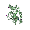
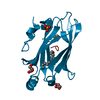
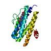
| Title | The structure of crystal form II of Haemophilus influenzae protein E | |||||||||
 Components Components | (PROTEIN E) x 2 | |||||||||
 Keywords Keywords | CELL ADHESION | |||||||||
| Function / homology |  Function and homology information Function and homology information | |||||||||
| Biological species |  HAEMOPHILUS INFLUENZAE (bacteria) HAEMOPHILUS INFLUENZAE (bacteria) | |||||||||
| Method |  X-RAY DIFFRACTION / SYNCHROTRON / MOLECULAR REPLACEMENT / Resolution: 2.102 Å X-RAY DIFFRACTION / SYNCHROTRON / MOLECULAR REPLACEMENT / Resolution: 2.102 Å | |||||||||
 Authors Authors | Singh, B. / Al-Jubair, T. / Riesbeck, K. / Thunnissen, M.M.G.M. | |||||||||
 Citation Citation | Journal: Infect.Immun. / Year: 2013 Title: The Unique Structure of Haemophilus Influenzae Protein E Reveals Multiple Binding Sites for Host Factors. Authors: Singh, B. / Al-Jubair, T. / Morgelin, M. / Thunnissen, M.M. / Riesbeck, K. | |||||||||
| History |
|
- Structure visualization
| Structure viewer | Molecule: MolmilJmol/JSmol |
|---|
- Downloads & links
-Download
| PDBx/mmCIF format | 3zh7.cif.gz | 165.4 KB | Display | PDBx/mmCIF format |
|---|---|---|---|---|
| PDB format | pdb3zh7.ent.gz | 134.3 KB | Display | PDB format |
| PDBx/mmJSON format | 3zh7.json.gz | Tree view | PDBx/mmJSON format | |
| Others |  Other downloads Other downloads |
-Validation report
| Arichive directory | https://data.pdbj.org/pub/pdb/validation_reports/zh/3zh7ftp://data.pdbj.org/pub/pdb/validation_reports/zh/3zh7 | HTTPS FTP |
|---|
-Related structure data


-Links
 PDBj
PDBj


- Assembly
| Deposited unit | 
| ||||||||
|---|---|---|---|---|---|---|---|---|---|
| 1 | 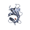
| ||||||||
| 2 | 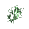
| ||||||||
| Unit cell |
| ||||||||
| Noncrystallographic symmetry (NCS) | NCS oper: (Code: given Matrix: (0.2825, -0.2293, -0.9315), Vector: |
-Components
| #1: Protein | Mass: 14719.706 Da / Num. of mol.: 1 Source method: isolated from a genetically manipulated source Source: (gene. exp.) HAEMOPHILUS INFLUENZAE (bacteria) / Production host: | ||||
|---|---|---|---|---|---|
| #2: Protein | Mass: 14861.797 Da / Num. of mol.: 1 Source method: isolated from a genetically manipulated source Source: (gene. exp.) HAEMOPHILUS INFLUENZAE (bacteria) / Production host: | ||||
| #3: Chemical |   Mass: 62.068 Da / Num. of mol.: 2 / Source method: obtained synthetically / Formula: C2H6O2 Mass: 62.068 Da / Num. of mol.: 2 / Source method: obtained synthetically / Formula: C2H6O2#4: Water | ChemComp-HOH / |  Mass: 18.015 Da / Num. of mol.: 47 / Source method: isolated from a natural source / Formula: H2O Mass: 18.015 Da / Num. of mol.: 47 / Source method: isolated from a natural source / Formula: H2OHas protein modification | Y | |
-Experimental details
-Experiment
| Experiment | Method: X-RAY DIFFRACTION / Number of used crystals: 1 |
|---|
- Sample preparation
| Crystal | Density Matthews: 1.75 Å3/Da / Density % sol: 29.7 % Description: STRUCTURE WAS SOLVED WITH STRUCTURE FROM OTHER CRYSTAL FORM OF PE. |
|---|---|
| Crystal grow | Details: 100 MM BISTRIS PROPANE PH 6.5, 200 MM NAI AND 20% W/V POLYETHYLENE GLYCOL 3350 |
-Data collection
| Diffraction | Mean temperature: 100 K |
|---|---|
| Diffraction source | Source: SYNCHROTRON / Site: MAX II  / Beamline: I911-3 / Wavelength: 1 / Beamline: I911-3 / Wavelength: 1 |
| Detector | Type: MARMOSAIC 225 mm CCD / Detector: CCD / Date: Jun 30, 2011 / Details: MIRRORS |
| Radiation | Monochromator: SI 111 / Protocol: SINGLE WAVELENGTH / Monochromatic (M) / Laue (L): M / Scattering type: x-ray |
| Radiation wavelength | Wavelength: 1 Å / Relative weight: 1 |
| Reflection | Resolution: 2.1→29 Å / Num. obs: 12867 / % possible obs: 91.4 % / Observed criterion σ(I): 2 / Redundancy: 2.49 % / Rmerge(I) obs: 0.06 / Net I/σ(I): 11.94 |
| Reflection shell | Highest resolution: 2.1 Å / Rmerge(I) obs: 0.46 / Mean I/σ(I) obs: 2.03 / % possible all: 79.8 |
- Processing
| Software |
| ||||||||||||||||||||||||||||||||||||||||||||||||||||||||||||||||||||||||||||||||||||||||||||||||||||||||||||||||||||||||||||||||||||||||||||||||||||||||||||||||||||||||||||||||||||||||||||||||||||||||
|---|---|---|---|---|---|---|---|---|---|---|---|---|---|---|---|---|---|---|---|---|---|---|---|---|---|---|---|---|---|---|---|---|---|---|---|---|---|---|---|---|---|---|---|---|---|---|---|---|---|---|---|---|---|---|---|---|---|---|---|---|---|---|---|---|---|---|---|---|---|---|---|---|---|---|---|---|---|---|---|---|---|---|---|---|---|---|---|---|---|---|---|---|---|---|---|---|---|---|---|---|---|---|---|---|---|---|---|---|---|---|---|---|---|---|---|---|---|---|---|---|---|---|---|---|---|---|---|---|---|---|---|---|---|---|---|---|---|---|---|---|---|---|---|---|---|---|---|---|---|---|---|---|---|---|---|---|---|---|---|---|---|---|---|---|---|---|---|---|---|---|---|---|---|---|---|---|---|---|---|---|---|---|---|---|---|---|---|---|---|---|---|---|---|---|---|---|---|---|---|---|---|
| Refinement | Method to determine structure: MOLECULAR REPLACEMENT / Resolution: 2.102→28.298 Å / SU ML: 0.57 / σ(F): 2.01 / Phase error: 27.47 / Stereochemistry target values: ML
| ||||||||||||||||||||||||||||||||||||||||||||||||||||||||||||||||||||||||||||||||||||||||||||||||||||||||||||||||||||||||||||||||||||||||||||||||||||||||||||||||||||||||||||||||||||||||||||||||||||||||
| Solvent computation | Shrinkage radii: 0.89 Å / VDW probe radii: 1 Å / Solvent model: FLAT BULK SOLVENT MODEL / Bsol: 40.955 Å2 / ksol: 0.439 e/Å3 | ||||||||||||||||||||||||||||||||||||||||||||||||||||||||||||||||||||||||||||||||||||||||||||||||||||||||||||||||||||||||||||||||||||||||||||||||||||||||||||||||||||||||||||||||||||||||||||||||||||||||
| Displacement parameters |
| ||||||||||||||||||||||||||||||||||||||||||||||||||||||||||||||||||||||||||||||||||||||||||||||||||||||||||||||||||||||||||||||||||||||||||||||||||||||||||||||||||||||||||||||||||||||||||||||||||||||||
| Refinement step | Cycle: LAST / Resolution: 2.102→28.298 Å
| ||||||||||||||||||||||||||||||||||||||||||||||||||||||||||||||||||||||||||||||||||||||||||||||||||||||||||||||||||||||||||||||||||||||||||||||||||||||||||||||||||||||||||||||||||||||||||||||||||||||||
| Refine LS restraints |
| ||||||||||||||||||||||||||||||||||||||||||||||||||||||||||||||||||||||||||||||||||||||||||||||||||||||||||||||||||||||||||||||||||||||||||||||||||||||||||||||||||||||||||||||||||||||||||||||||||||||||
| LS refinement shell |
| ||||||||||||||||||||||||||||||||||||||||||||||||||||||||||||||||||||||||||||||||||||||||||||||||||||||||||||||||||||||||||||||||||||||||||||||||||||||||||||||||||||||||||||||||||||||||||||||||||||||||
| Refinement TLS params. | Method: refined / Refine-ID: X-RAY DIFFRACTION
| ||||||||||||||||||||||||||||||||||||||||||||||||||||||||||||||||||||||||||||||||||||||||||||||||||||||||||||||||||||||||||||||||||||||||||||||||||||||||||||||||||||||||||||||||||||||||||||||||||||||||
| Refinement TLS group |
|
