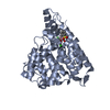
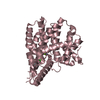
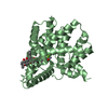
Entry Database : PDB / ID : 3gwtTitle Catalytic domain of human phosphodiesterase 4B2B in complex with a quinoline inhibitor cAMP-specific 3',5'-cyclic phosphodiesterase 4B Keywords / / / Function / homology Function Domain/homology Component
/ / / / / / / / / / / / / / / / / / / / / / / / / / / / / / / / / / / / / / / / / / / / / / / / / / / / / / / / / / / / / / / / Biological species Homo sapiens (human)Method / / / Resolution : 1.75 Å Authors Somers, D.O. / Neu, M. Journal : Bioorg.Med.Chem.Lett. / Year : 2009Title : Quinolines as a novel structural class of potent and selective PDE4 inhibitors. Optimisation for inhaled administration.Authors: Woodrow, M.D. / Ballantine, S.P. / Barker, M.D. / Clarke, B.J. / Dawson, J. / Dean, T.W. / Delves, C.J. / Evans, B. / Gough, S.L. / Guntrip, S.B. / Holman, S. / Holmes, D.S. / Kranz, M. / ... Authors : Woodrow, M.D. / Ballantine, S.P. / Barker, M.D. / Clarke, B.J. / Dawson, J. / Dean, T.W. / Delves, C.J. / Evans, B. / Gough, S.L. / Guntrip, S.B. / Holman, S. / Holmes, D.S. / Kranz, M. / Lindvaal, M.K. / Lucas, F.S. / Neu, M. / Ranshaw, L.E. / Solanke, Y.E. / Somers, D.O. / Ward, P. / Wiseman, J.O. History Deposition Apr 1, 2009 Deposition site / Processing site Revision 1.0 Apr 7, 2010 Provider / Type Revision 1.1 Jul 13, 2011 Group Revision 1.2 Nov 1, 2017 Group / Category Revision 1.3 Oct 13, 2021 Group / Derived calculationsCategory database_2 / pdbx_struct_conn_angle ... database_2 / pdbx_struct_conn_angle / struct_conn / struct_ref_seq_dif / struct_site Item _database_2.pdbx_DOI / _database_2.pdbx_database_accession ... _database_2.pdbx_DOI / _database_2.pdbx_database_accession / _pdbx_struct_conn_angle.ptnr1_auth_comp_id / _pdbx_struct_conn_angle.ptnr1_auth_seq_id / _pdbx_struct_conn_angle.ptnr1_label_asym_id / _pdbx_struct_conn_angle.ptnr1_label_atom_id / _pdbx_struct_conn_angle.ptnr1_label_comp_id / _pdbx_struct_conn_angle.ptnr1_label_seq_id / _pdbx_struct_conn_angle.ptnr2_auth_comp_id / _pdbx_struct_conn_angle.ptnr2_auth_seq_id / _pdbx_struct_conn_angle.ptnr2_label_asym_id / _pdbx_struct_conn_angle.ptnr2_label_atom_id / _pdbx_struct_conn_angle.ptnr2_label_comp_id / _pdbx_struct_conn_angle.ptnr3_auth_comp_id / _pdbx_struct_conn_angle.ptnr3_auth_seq_id / _pdbx_struct_conn_angle.ptnr3_label_asym_id / _pdbx_struct_conn_angle.ptnr3_label_atom_id / _pdbx_struct_conn_angle.ptnr3_label_comp_id / _pdbx_struct_conn_angle.ptnr3_label_seq_id / _pdbx_struct_conn_angle.value / _struct_conn.pdbx_dist_value / _struct_conn.ptnr1_auth_comp_id / _struct_conn.ptnr1_auth_seq_id / _struct_conn.ptnr1_label_asym_id / _struct_conn.ptnr1_label_atom_id / _struct_conn.ptnr1_label_comp_id / _struct_conn.ptnr1_label_seq_id / _struct_conn.ptnr2_auth_comp_id / _struct_conn.ptnr2_auth_seq_id / _struct_conn.ptnr2_label_asym_id / _struct_conn.ptnr2_label_atom_id / _struct_conn.ptnr2_label_comp_id / _struct_ref_seq_dif.details / _struct_site.pdbx_auth_asym_id / _struct_site.pdbx_auth_comp_id / _struct_site.pdbx_auth_seq_id Revision 1.4 Sep 6, 2023 Group / Refinement descriptionCategory / chem_comp_bond / pdbx_initial_refinement_model
Show all Show less
 Movie
Movie Controller

 Yorodumi
 Open data
 Basic information
 Components
 Keywords
 Function and homology information
 Homo sapiens (human)
 X-RAY DIFFRACTION /
  Authors
 Citation
 Structure visualization
 Downloads & links
 Other downloads
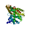

 PDBj
PDBj


 Assembly


 Spodoptera frugiperda (fall armyworm)


 Mass: 518.584 Da / Num. of mol.: 1 / Source method: obtained synthetically / Formula: C27H26N4O5S
 Mass: 65.409 Da / Num. of mol.: 1 / Source method: obtained synthetically / Formula: Zn
 Mass: 24.305 Da / Num. of mol.: 1 / Source method: obtained synthetically / Formula: Mg
 Mass: 74.922 Da / Num. of mol.: 4 / Source method: obtained synthetically / Formula: As
 Mass: 92.094 Da / Num. of mol.: 3 / Source method: obtained synthetically / Formula: C3H8O3
 Sample preparation
 / Beamline: ID14-4 / Wavelength: 0.9393 Å
 Processing