 Movie
Movie Controller

[English] 日本語
 Yorodumi
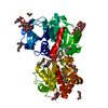
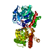
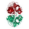
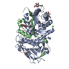
- PDB-3guh: Crystal Structure of Wild-type E.coli GS in complex with ADP and DGM -
+ Open data

- Basic information
| Entry | Database: PDB / ID: 3guh | ||||||
|---|---|---|---|---|---|---|---|
| Title | Crystal Structure of Wild-type E.coli GS in complex with ADP and DGM | ||||||
 Components Components | Glycogen synthase | ||||||
 Keywords Keywords | TRANSFERASE / glycosyl-transferase / GT-B fold / Rossmann fold / closed-form / ADP and intermediate D-glucopyranosylium binding / Glycogen biosynthesis / Glycosyltransferase | ||||||
| Function / homology |  Function and homology information Function and homology informationstarch synthase (glycosyl-transferring) / alpha-1,4-glucan glucosyltransferase (ADP-glucose donor) activity / alpha-1,4-glucan glucosyltransferase (UDP-glucose donor) activity / glycogen biosynthetic process / DNA damage response / cytosol Similarity search - Function | ||||||
| Biological species |  | ||||||
| Method |  X-RAY DIFFRACTION / SYNCHROTRON / MOLECULAR REPLACEMENT / molecular replacement / Resolution: 2.79 Å X-RAY DIFFRACTION / SYNCHROTRON / MOLECULAR REPLACEMENT / molecular replacement / Resolution: 2.79 Å | ||||||
 Authors Authors | Sheng, F. / Geiger, J. | ||||||
 Citation Citation | Journal: J.Biol.Chem. / Year: 2009 Title: The Crystal Structures of the Open and Catalytically Competent Closed Conformation of Escherichia coli Glycogen Synthase. Authors: Sheng, F. / Jia, X. / Yep, A. / Preiss, J. / Geiger, J.H. | ||||||
| History |
|
- Structure visualization
| Structure viewer | Molecule: MolmilJmol/JSmol |
|---|
- Downloads & links
-Download
| PDBx/mmCIF format | 3guh.cif.gz | 117.4 KB | Display | PDBx/mmCIF format |
|---|---|---|---|---|
| PDB format | pdb3guh.ent.gz | 87.1 KB | Display | PDB format |
| PDBx/mmJSON format | 3guh.json.gz | Tree view | PDBx/mmJSON format | |
| Others |  Other downloads Other downloads |
-Validation report
| Arichive directory | https://data.pdbj.org/pub/pdb/validation_reports/gu/3guhftp://data.pdbj.org/pub/pdb/validation_reports/gu/3guh | HTTPS FTP |
|---|
-Related structure data
| Related structure data |  2qzsC 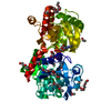 2r4tC  2r4uC 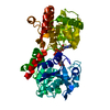 3copC  3d1jC  1rzuS  2qyy C: citing same article ( S: Starting model for refinement |
|---|---|
| Similar structure data |


-Links
 PDBj
PDBj
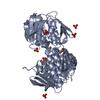
- Assembly
| Deposited unit | 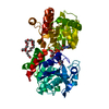
| ||||||||
|---|---|---|---|---|---|---|---|---|---|
| 1 |
| ||||||||
| Unit cell |
|
-Components
-Protein / Sugars , 2 types, 2 molecules A
| #1: Protein | Mass: 53951.348 Da / Num. of mol.: 1 Source method: isolated from a genetically manipulated source Source: (gene. exp.) References: UniProt: P0A6U8, starch synthase (glycosyl-transferring) |
|---|---|
| #4: Sugar | ChemComp-ASO /  Type: D-saccharide / Mass: 164.156 Da / Num. of mol.: 1 Type: D-saccharide / Mass: 164.156 Da / Num. of mol.: 1Source method: isolated from a genetically manipulated source Formula: C6H12O5 |
-Non-polymers , 5 types, 215 molecules 


| #2: Chemical | ChemComp-ADP /  Mass: 427.201 Da / Num. of mol.: 1 / Source method: obtained synthetically / Formula: C10H15N5O10P2 / Comment: ADP, energy-carrying molecule*YM Mass: 427.201 Da / Num. of mol.: 1 / Source method: obtained synthetically / Formula: C10H15N5O10P2 / Comment: ADP, energy-carrying molecule*YM | ||||
|---|---|---|---|---|---|
| #3: Chemical | ChemComp-250 / ( Mass: 268.331 Da / Num. of mol.: 1 / Source method: obtained synthetically / Formula: C9H20N2O5S Mass: 268.331 Da / Num. of mol.: 1 / Source method: obtained synthetically / Formula: C9H20N2O5S | ||||
| #5: Chemical |  Mass: 634.751 Da / Num. of mol.: 2 / Source method: obtained synthetically / Formula: C28H58O15 Mass: 634.751 Da / Num. of mol.: 2 / Source method: obtained synthetically / Formula: C28H58O15#6: Chemical |  Mass: 59.044 Da / Num. of mol.: 2 / Source method: obtained synthetically / Formula: C2H3O2 Mass: 59.044 Da / Num. of mol.: 2 / Source method: obtained synthetically / Formula: C2H3O2#7: Water | ChemComp-HOH / | Mass: 18.015 Da / Num. of mol.: 209 / Source method: isolated from a natural source / Formula: H2O |
-Details
| Nonpolymer details | THE ASO SPECIES IN THIS PDB ENTRY DOES NOT HAVE HYDROGEN ATOM ON C1, THEREFORE IT IS POSITIVELY ...THE ASO SPECIES IN THIS PDB ENTRY DOES NOT HAVE HYDROGEN ATOM ON C1, THEREFORE IT IS POSITIVELY |
|---|
-Experimental details
-Experiment
| Experiment | Method: X-RAY DIFFRACTION / Number of used crystals: 1 |
|---|
- Sample preparation
| Crystal | Density Matthews: 5.62 Å3/Da / Density % sol: 78.13 % |
|---|---|
| Crystal grow | Temperature: 277 K / Method: vapor diffusion, hanging drop / pH: 8.1 Details: 40% (w/v) PEG 4000, 0.2 M NaAc, 0.1 M HEPPSO, pH 8.1, VAPOR DIFFUSION, HANGING DROP, temperature 277K |
-Data collection
| Diffraction | Mean temperature: 98 K |
|---|---|
| Diffraction source | Source: SYNCHROTRON / Site: APS  / Beamline: 14-ID-B / Wavelength: 1 Å / Beamline: 14-ID-B / Wavelength: 1 Å |
| Detector | Type: MAR CCD 165 mm / Detector: CCD / Date: May 25, 2005 |
| Radiation | Protocol: SINGLE WAVELENGTH / Monochromatic (M) / Laue (L): M / Scattering type: x-ray |
| Radiation wavelength | Wavelength: 1 Å / Relative weight: 1 |
| Reflection | Resolution: 2.79→97.13 Å / Num. all: 29589 / Num. obs: 29543 / % possible obs: 99.2 % / Observed criterion σ(F): 2 / Observed criterion σ(I): 1.91 / Redundancy: 8.1 % / Biso Wilson estimate: 62.3 Å2 / Rmerge(I) obs: 0.146 / Χ2: 1.976 / Net I/σ(I): 23.996 |
| Reflection shell | Resolution: 2.79→2.9 Å / Redundancy: 6.1 % / Rmerge(I) obs: 0.516 / Num. unique all: 2740 / Χ2: 0.511 / % possible all: 92.9 |
-Phasing
| Phasing | Method: molecular replacement | |||||||||
|---|---|---|---|---|---|---|---|---|---|---|
| Phasing MR | Rfactor: 0.349 / Cor.coef. Fo:Fc: 0.744
|
- Processing
| Software |
| |||||||||||||||||||||||||||||||||||||||||||||||||||||||||||||||||
|---|---|---|---|---|---|---|---|---|---|---|---|---|---|---|---|---|---|---|---|---|---|---|---|---|---|---|---|---|---|---|---|---|---|---|---|---|---|---|---|---|---|---|---|---|---|---|---|---|---|---|---|---|---|---|---|---|---|---|---|---|---|---|---|---|---|---|
| Refinement | Method to determine structure: MOLECULAR REPLACEMENT Starting model: PDB ENTRY 1RZU Resolution: 2.79→50 Å / Cor.coef. Fo:Fc: 0.954 / Cor.coef. Fo:Fc free: 0.938 / WRfactor Rfree: 0.184 / WRfactor Rwork: 0.156 / Occupancy max: 1 / Occupancy min: 0.2 / FOM work R set: 0.861 / SU B: 7.902 / SU ML: 0.154 / SU R Cruickshank DPI: 0.287 / SU Rfree: 0.219 / Cross valid method: THROUGHOUT / σ(F): 0 / ESU R: 0.287 / ESU R Free: 0.219 / Stereochemistry target values: MAXIMUM LIKELIHOOD / Details: HYDROGENS HAVE BEEN ADDED IN THE RIDING POSITIONS
| |||||||||||||||||||||||||||||||||||||||||||||||||||||||||||||||||
| Solvent computation | Ion probe radii: 0.8 Å / Shrinkage radii: 0.8 Å / VDW probe radii: 1.4 Å / Solvent model: MASK | |||||||||||||||||||||||||||||||||||||||||||||||||||||||||||||||||
| Displacement parameters | Biso max: 122.93 Å2 / Biso mean: 40.525 Å2 / Biso min: 2 Å2
| |||||||||||||||||||||||||||||||||||||||||||||||||||||||||||||||||
| Refinement step | Cycle: LAST / Resolution: 2.79→50 Å
| |||||||||||||||||||||||||||||||||||||||||||||||||||||||||||||||||
| Refine LS restraints |
| |||||||||||||||||||||||||||||||||||||||||||||||||||||||||||||||||
| LS refinement shell | Resolution: 2.79→2.864 Å / Total num. of bins used: 20
|
