 Movie
Movie Controller

[English] 日本語
 Yorodumi
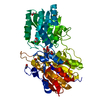
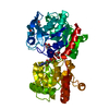
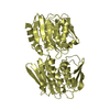
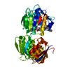
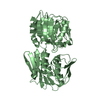
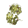
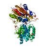
- PDB-2r4t: Crystal Structure of Wild-type E.coli GS in Complex with ADP and ... -
+ Open data

- Basic information
| Entry | Database: PDB / ID: 2r4t | ||||||
|---|---|---|---|---|---|---|---|
| Title | Crystal Structure of Wild-type E.coli GS in Complex with ADP and Glucose(wtGSc) | ||||||
 Components Components | Glycogen synthase | ||||||
 Keywords Keywords | TRANSFERASE / glycosyl-transferase / GT-B fold / Rossmann fold / closed-form / ADP and glucose binding | ||||||
| Function / homology |  Function and homology information Function and homology informationstarch synthase (glycosyl-transferring) / alpha-1,4-glucan glucosyltransferase (ADP-glucose donor) activity / alpha-1,4-glucan glucosyltransferase (UDP-glucose donor) activity / glycogen biosynthetic process / DNA damage response / cytosol Similarity search - Function | ||||||
| Biological species |  | ||||||
| Method |  X-RAY DIFFRACTION / SYNCHROTRON / MOLECULAR REPLACEMENT / molecular replacement / Resolution: 2.258 Å X-RAY DIFFRACTION / SYNCHROTRON / MOLECULAR REPLACEMENT / molecular replacement / Resolution: 2.258 Å | ||||||
 Authors Authors | Sheng, F. / Geiger, J. | ||||||
 Citation Citation | Journal: J.Biol.Chem. / Year: 2009 Title: The crystal structures of the open and catalytically competent closed conformation of Escherichia coli glycogen synthase. Authors: Sheng, F. / Jia, X. / Yep, A. / Preiss, J. / Geiger, J.H. | ||||||
| History |
|
- Structure visualization
| Structure viewer | Molecule: MolmilJmol/JSmol |
|---|
- Downloads & links
-Download
| PDBx/mmCIF format | 2r4t.cif.gz | 124.4 KB | Display | PDBx/mmCIF format |
|---|---|---|---|---|
| PDB format | pdb2r4t.ent.gz | 89.6 KB | Display | PDB format |
| PDBx/mmJSON format | 2r4t.json.gz | Tree view | PDBx/mmJSON format | |
| Others |  Other downloads Other downloads |
-Validation report
| Arichive directory | https://data.pdbj.org/pub/pdb/validation_reports/r4/2r4tftp://data.pdbj.org/pub/pdb/validation_reports/r4/2r4t | HTTPS FTP |
|---|
-Related structure data
| Related structure data |  2qzsSC  2r4uC 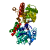 3copC  3d1jC 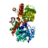 3guhC  2qyy S: Starting model for refinement C: citing same article ( |
|---|---|
| Similar structure data |


-Links
 PDBj
PDBj


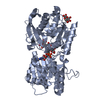
- Assembly
| Deposited unit | 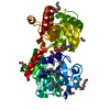
| ||||||||
|---|---|---|---|---|---|---|---|---|---|
| 1 |
| ||||||||
| Unit cell |
|
-Components
-Protein / Sugars , 2 types, 2 molecules A
| #1: Protein | Mass: 53951.348 Da / Num. of mol.: 1 Source method: isolated from a genetically manipulated source Source: (gene. exp.) References: UniProt: P0A6U8, starch synthase (glycosyl-transferring) |
|---|---|
| #2: Sugar | ChemComp-GLC /  Type: D-saccharide, alpha linking / Mass: 180.156 Da / Num. of mol.: 1 Type: D-saccharide, alpha linking / Mass: 180.156 Da / Num. of mol.: 1Source method: isolated from a genetically manipulated source Formula: C6H12O6 |
-Non-polymers , 4 types, 350 molecules 


| #3: Chemical | ChemComp-ADP /  Mass: 427.201 Da / Num. of mol.: 1 / Source method: obtained synthetically / Formula: C10H15N5O10P2 / Comment: ADP, energy-carrying molecule*YM Mass: 427.201 Da / Num. of mol.: 1 / Source method: obtained synthetically / Formula: C10H15N5O10P2 / Comment: ADP, energy-carrying molecule*YM | ||
|---|---|---|---|
| #4: Chemical | ChemComp-250 / ( Mass: 268.331 Da / Num. of mol.: 1 / Source method: obtained synthetically / Formula: C9H20N2O5S Mass: 268.331 Da / Num. of mol.: 1 / Source method: obtained synthetically / Formula: C9H20N2O5S | ||
| #5: Chemical | ChemComp-PE3 /  Mass: 634.751 Da / Num. of mol.: 13 / Source method: obtained synthetically / Formula: C28H58O15 Mass: 634.751 Da / Num. of mol.: 13 / Source method: obtained synthetically / Formula: C28H58O15#6: Water | ChemComp-HOH / | Mass: 18.015 Da / Num. of mol.: 335 / Source method: isolated from a natural source / Formula: H2O |
-Experimental details
-Experiment
| Experiment | Method: X-RAY DIFFRACTION / Number of used crystals: 1 |
|---|
- Sample preparation
| Crystal | Density Matthews: 5.7 Å3/Da / Density % sol: 78.3 % |
|---|---|
| Crystal grow | Temperature: 277 K / Method: vapor diffusion, hanging drop / pH: 7.6 Details: 40%(w/v) PEG 4000, 0.2 M Na tartrate and 0.1 M HEPPSO (pH 7.6), VAPOR DIFFUSION, HANGING DROP, temperature 277K |
-Data collection
| Diffraction | Mean temperature: 100 K |
|---|---|
| Diffraction source | Source: SYNCHROTRON / Site: APS  / Beamline: 5ID-B / Wavelength: 1 Å / Beamline: 5ID-B / Wavelength: 1 Å |
| Detector | Type: MARMOSAIC 225 mm CCD / Detector: CCD / Date: Jul 24, 2006 |
| Radiation | Monochromator: graphite / Protocol: SINGLE WAVELENGTH / Monochromatic (M) / Laue (L): M / Scattering type: x-ray |
| Radiation wavelength | Wavelength: 1 Å / Relative weight: 1 |
| Reflection | Resolution: 2.258→97.13 Å / Num. obs: 56077 / % possible obs: 99.9 % / Observed criterion σ(I): 4.5 / Redundancy: 5.1 % / Biso Wilson estimate: 41.1 Å2 / Rmerge(I) obs: 0.052 / Χ2: 1.001 / Net I/σ(I): 11.8 |
| Reflection shell | Resolution: 2.258→2.34 Å / Redundancy: 4.9 % / Rmerge(I) obs: 0.287 / Mean I/σ(I) obs: 4.5 / Num. unique all: 5555 / Χ2: 0.635 / % possible all: 99.8 |
-Phasing
| Phasing | Method: molecular replacement | |||||||||
|---|---|---|---|---|---|---|---|---|---|---|
| Phasing MR | Rfactor: 0.306 / Cor.coef. Fo:Fc: 0.77
|
- Processing
| Software |
| ||||||||||||||||||||||||||||||||||||||||||||||||||||||||||||||||||||||||||||||||||||||||||
|---|---|---|---|---|---|---|---|---|---|---|---|---|---|---|---|---|---|---|---|---|---|---|---|---|---|---|---|---|---|---|---|---|---|---|---|---|---|---|---|---|---|---|---|---|---|---|---|---|---|---|---|---|---|---|---|---|---|---|---|---|---|---|---|---|---|---|---|---|---|---|---|---|---|---|---|---|---|---|---|---|---|---|---|---|---|---|---|---|---|---|---|
| Refinement | Method to determine structure: MOLECULAR REPLACEMENT Starting model: 2QZS Resolution: 2.258→97.13 Å / Cor.coef. Fo:Fc: 0.965 / Cor.coef. Fo:Fc free: 0.959 / SU B: 2.944 / SU ML: 0.074 / Cross valid method: THROUGHOUT / σ(I): 4.5 / ESU R: 0.127 / ESU R Free: 0.116 / Stereochemistry target values: MAXIMUM LIKELIHOOD / Details: HYDROGENS HAVE BEEN ADDED IN THE RIDING POSITIONS
| ||||||||||||||||||||||||||||||||||||||||||||||||||||||||||||||||||||||||||||||||||||||||||
| Solvent computation | Ion probe radii: 0.8 Å / Shrinkage radii: 0.8 Å / VDW probe radii: 1.2 Å / Solvent model: BABINET MODEL WITH MASK | ||||||||||||||||||||||||||||||||||||||||||||||||||||||||||||||||||||||||||||||||||||||||||
| Displacement parameters | Biso mean: 37.998 Å2
| ||||||||||||||||||||||||||||||||||||||||||||||||||||||||||||||||||||||||||||||||||||||||||
| Refinement step | Cycle: LAST / Resolution: 2.258→97.13 Å
| ||||||||||||||||||||||||||||||||||||||||||||||||||||||||||||||||||||||||||||||||||||||||||
| Refine LS restraints |
| ||||||||||||||||||||||||||||||||||||||||||||||||||||||||||||||||||||||||||||||||||||||||||
| LS refinement shell | Resolution: 2.258→2.317 Å / Total num. of bins used: 20
|
