 Movie
Movie Controller

[English] 日本語
 Yorodumi
- PDB-1rf5: Structural Studies of Streptococcus pneumoniae EPSP Synthase in U... -
+ Open data

- Basic information
| Entry | Database: PDB / ID: 1rf5 | ||||||
|---|---|---|---|---|---|---|---|
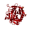
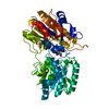
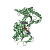
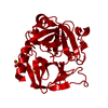
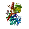
| Title | Structural Studies of Streptococcus pneumoniae EPSP Synthase in Unliganded State | ||||||
 Components Components | 5-enolpyruvylshikimate-3-phosphate synthase | ||||||
 Keywords Keywords | TRANSFERASE / shikimate pathway / EPSP synthase / S3P / Glyphosate / PEP / S. pneumoniae | ||||||
| Function / homology |  Function and homology information Function and homology information3-phosphoshikimate 1-carboxyvinyltransferase / 3-phosphoshikimate 1-carboxyvinyltransferase activity / chorismate biosynthetic process / aromatic amino acid biosynthetic process / amino acid biosynthetic process / cytoplasm Similarity search - Function | ||||||
| Biological species |   Streptococcus pneumoniae (bacteria) Streptococcus pneumoniae (bacteria) | ||||||
| Method |  X-RAY DIFFRACTION / SYNCHROTRON / MAD / Resolution: 2.3 Å X-RAY DIFFRACTION / SYNCHROTRON / MAD / Resolution: 2.3 Å | ||||||
 Authors Authors | Park, H. / Hilsenbeck, J.L. / Kim, H.J. / Shuttleworth, W.A. / Park, Y.H. / Evans, J.N. / Kang, C. | ||||||
 Citation Citation | Journal: Mol.Microbiol. / Year: 2004 Title: Structural studies of Streptococcus pneumoniae EPSP synthase in unliganded state, tetrahedral intermediate-bound state and S3P-GLP-bound state. Authors: Park, H. / Hilsenbeck, J.L. / Kim, H.J. / Shuttleworth, W.A. / Park, Y.H. / Evans, J.N. / Kang, C. | ||||||
| History |
|
- Structure visualization
| Structure viewer | Molecule: MolmilJmol/JSmol |
|---|
- Downloads & links
-Download
| PDBx/mmCIF format | 1rf5.cif.gz | 334.9 KB | Display | PDBx/mmCIF format |
|---|---|---|---|---|
| PDB format | pdb1rf5.ent.gz | 274.7 KB | Display | PDB format |
| PDBx/mmJSON format | 1rf5.json.gz | Tree view | PDBx/mmJSON format | |
| Others |  Other downloads Other downloads |
-Validation report
| Arichive directory | https://data.pdbj.org/pub/pdb/validation_reports/rf/1rf5ftp://data.pdbj.org/pub/pdb/validation_reports/rf/1rf5 | HTTPS FTP |
|---|
-Related structure data


-Links
 PDBj
PDBj
- Assembly
| Deposited unit | 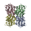
| ||||||||
|---|---|---|---|---|---|---|---|---|---|
| 1 | 
| ||||||||
| 2 | 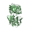
| ||||||||
| 3 | 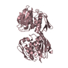
| ||||||||
| 4 | 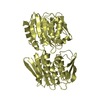
| ||||||||
| Unit cell |
| ||||||||
| Details | The biological assembly is a monomer |
-Components
| #1: Protein | Mass: 45878.820 Da / Num. of mol.: 4 Source method: isolated from a genetically manipulated source Source: (gene. exp.) Streptococcus pneumoniae (bacteria) / Gene: aroA / Production host: References: UniProt: Q9S400, 3-phosphoshikimate 1-carboxyvinyltransferase #2: Water | ChemComp-HOH / |  Mass: 18.015 Da / Num. of mol.: 767 / Source method: isolated from a natural source / Formula: H2O Mass: 18.015 Da / Num. of mol.: 767 / Source method: isolated from a natural source / Formula: H2O |
|---|
-Experimental details
-Experiment
| Experiment | Method: X-RAY DIFFRACTION / Number of used crystals: 1 |
|---|
- Sample preparation
| Crystal | Density Matthews: 3.23 Å3/Da / Density % sol: 61.95 % | |||||||||||||||||||||||||||||||||||||||||||||||||
|---|---|---|---|---|---|---|---|---|---|---|---|---|---|---|---|---|---|---|---|---|---|---|---|---|---|---|---|---|---|---|---|---|---|---|---|---|---|---|---|---|---|---|---|---|---|---|---|---|---|---|
| Crystal grow | Temperature: 277 K / Method: vapor diffusion, hanging drop / pH: 7.5 Details: ammonium sulfate, NaCl, HEPES, pH 7.5, VAPOR DIFFUSION, HANGING DROP, temperature 277K | |||||||||||||||||||||||||||||||||||||||||||||||||
| Crystal grow | *PLUS Temperature: 4 ℃ / Method: vapor diffusion, hanging drop | |||||||||||||||||||||||||||||||||||||||||||||||||
| Components of the solutions | *PLUS
|
-Data collection
| Diffraction | Mean temperature: 100 K | ||||||||||||
|---|---|---|---|---|---|---|---|---|---|---|---|---|---|
| Diffraction source | Source: SYNCHROTRON / Site: SSRL  / Beamline: BL9-2 / Wavelength: 0.992, 0.980, 0.9183 / Beamline: BL9-2 / Wavelength: 0.992, 0.980, 0.9183 | ||||||||||||
| Detector | Type: ADSC QUANTUM 315 / Detector: CCD / Date: Dec 5, 2001 | ||||||||||||
| Radiation | Monochromator: GRAPHITE / Protocol: MAD / Monochromatic (M) / Laue (L): M / Scattering type: x-ray | ||||||||||||
| Radiation wavelength |
| ||||||||||||
| Reflection | Resolution: 2.2→30 Å / Num. all: 102641 / Num. obs: 102641 / % possible obs: 97.2 % / Observed criterion σ(F): 1 / Observed criterion σ(I): 1 | ||||||||||||
| Reflection shell | Resolution: 2.2→2.3 Å / % possible all: 96.5 | ||||||||||||
| Reflection | *PLUS Num. measured all: 982621 / Rmerge(I) obs: 0.036 | ||||||||||||
| Reflection shell | *PLUS % possible obs: 96.5 % / Rmerge(I) obs: 0.128 |
- Processing
| Software |
| ||||||||||||||||||||
|---|---|---|---|---|---|---|---|---|---|---|---|---|---|---|---|---|---|---|---|---|---|
| Refinement | Method to determine structure: MAD / Resolution: 2.3→10 Å / σ(F): 2 / Stereochemistry target values: Engh & Huber Details: The peptide bond distance between GLU 340 and THR 341 of chains A and C, and between GLU 334 and GLU 335 of chain C, are slightly longer than typical peptide bond distance. Residues 335-340 ...Details: The peptide bond distance between GLU 340 and THR 341 of chains A and C, and between GLU 334 and GLU 335 of chain C, are slightly longer than typical peptide bond distance. Residues 335-340 were set to 0 occupancy and was not included in refinement.
| ||||||||||||||||||||
| Refinement step | Cycle: LAST / Resolution: 2.3→10 Å
| ||||||||||||||||||||
| Refine LS restraints |
| ||||||||||||||||||||
| Refinement | *PLUS Num. reflection all: 95252 / % reflection Rfree: 5 % / Rfactor Rfree: 0.254 | ||||||||||||||||||||
| Solvent computation | *PLUS | ||||||||||||||||||||
| Displacement parameters | *PLUS | ||||||||||||||||||||
| Refine LS restraints | *PLUS Type: x_angle_deg / Dev ideal: 2.49 | ||||||||||||||||||||
| LS refinement shell | *PLUS Rfactor Rfree: 0.272 / Rfactor Rwork: 0.219 |
