- PDB-3grz: CRYSTAL STRUCTURE OF ribosomal protein L11 methylase FROM Lactoba... -
+
Open data
ID or keywords:
Loading...
-
Basic information
Entry
Database: PDB / ID: 3grz
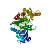
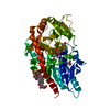
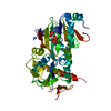
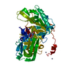
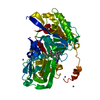
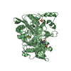
Title
CRYSTAL STRUCTURE OF ribosomal protein L11 methylase FROM Lactobacillus delbrueckii subsp. bulgaricus
Components
Ribosomal protein L11 methyltransferase
Keywords
TRANSFERASE / METHYLASE / SAM-BINDING DOMAIN / PSI-2 / NYSGXRC / STRUCTURAL GENOMICS / PROTEIN STRUCTURE INITIATIVE / NEW YORK SGX RESEARCH CENTER FOR STRUCTURAL GENOMICS / Methyltransferase
Single alpha-helices involved in coiled-coils or other helix-helix interfaces - #830 / Ribosomal protein L11 methyltransferase / : / Ribosomal protein L11 methyltransferase (PrmA) / Single alpha-helices involved in coiled-coils or other helix-helix interfaces / Vaccinia Virus protein VP39 / Helix non-globular / Special / S-adenosyl-L-methionine-dependent methyltransferase superfamily / Rossmann fold ...Single alpha-helices involved in coiled-coils or other helix-helix interfaces - #830 / Ribosomal protein L11 methyltransferase / : / Ribosomal protein L11 methyltransferase (PrmA) / Single alpha-helices involved in coiled-coils or other helix-helix interfaces / Vaccinia Virus protein VP39 / Helix non-globular / Special / S-adenosyl-L-methionine-dependent methyltransferase superfamily / Rossmann fold / 3-Layer(aba) Sandwich / Alpha Beta Similarity search - Domain/homology
Mass: 18.015 Da / Num. of mol.: 233 / Source method: isolated from a natural source / Formula: H2O
Sequence details
THE TARGET WAS EXPRESSED AS A FULL-LENGTH PROTEIN HOWEVER THE N-TERMINAL DOMAIN THAT INCLUDES ...THE TARGET WAS EXPRESSED AS A FULL-LENGTH PROTEIN HOWEVER THE N-TERMINAL DOMAIN THAT INCLUDES RESIDUES FROM 3 TO ABOUT 116 WAS LOST DURING CRYSTALLIZATION LIKELY DUE TO PROTEOLYSIS
-
Experimental details
-
Experiment
Experiment
Method: X-RAY DIFFRACTION / Number of used crystals: 1
-
Sample preparation
Crystal
Density Matthews: 2.5 Å3/Da / Density % sol: 50.84 %
Crystal grow
Temperature: 294 K / Method: vapor diffusion, sitting drop / pH: 6.2 Details: 0.1M SODIUM-POTASSIUM PHOSPHATE PH 6. 10% PEG3K, 10% GLYCEROL, VAPOR DIFFUSION, SITTING DROP, TEMPERATURE 294K
Protocol: SINGLE WAVELENGTH / Monochromatic (M) / Laue (L): M / Scattering type: x-ray
Radiation wavelength
Wavelength: 0.9791 Å / Relative weight: 1
Reflection
Resolution: 2→50 Å / Num. obs: 30326 / % possible obs: 98.1 % / Observed criterion σ(I): -0.5 / Redundancy: 2.1 % / Biso Wilson estimate: 36.872 Å2 / Rsym value: 0.051 / Net I/σ(I): 9.5
Reflection shell
Resolution: 2→2.07 Å / Redundancy: 2 % / Rmerge(I) obs: 0.62 / Mean I/σ(I) obs: 1.1 / % possible all: 90.6
-
Processing
Software
Name
Version
Classification
SHELXD
phasing
REFMAC
5.3.0034
refinement
DENZO
datareduction
HKL-2000
datascaling
Refinement
Method to determine structure: SAD / Resolution: 2→20 Å / Cor.coef. Fo:Fc: 0.969 / Cor.coef. Fo:Fc free: 0.946 / SU B: 4.248 / SU ML: 0.118 / Cross valid method: THROUGHOUT / ESU R: 0.162 / ESU R Free: 0.154 / Stereochemistry target values: MAXIMUM LIKELIHOOD / Details: HYDROGENS HAVE BEEN ADDED IN THE RIDING POSITIONS
Rfactor
Num. reflection
% reflection
Selection details
Rfree
0.22461
965
3.2 %
RANDOM
Rwork
0.17356
-
-
-
obs
0.17523
28941
98.76 %
-
Solvent computation
Ion probe radii: 0.8 Å / Shrinkage radii: 0.8 Å / VDW probe radii: 1.4 Å / Solvent model: MASK
In the structure databanks used in Yorodumi, some data are registered as the other names, "COVID-19 virus" and "2019-nCoV". Here are the details of the virus and the list of structure data.
Jan 31, 2019. EMDB accession codes are about to change! (news from PDBe EMDB page)
EMDB accession codes are about to change! (news from PDBe EMDB page)
The allocation of 4 digits for EMDB accession codes will soon come to an end. Whilst these codes will remain in use, new EMDB accession codes will include an additional digit and will expand incrementally as the available range of codes is exhausted. The current 4-digit format prefixed with “EMD-” (i.e. EMD-XXXX) will advance to a 5-digit format (i.e. EMD-XXXXX), and so on. It is currently estimated that the 4-digit codes will be depleted around Spring 2019, at which point the 5-digit format will come into force.
The EM Navigator/Yorodumi systems omit the EMD- prefix.
Related info.:Q: What is EMD? / ID/Accession-code notation in Yorodumi/EM Navigator
Yorodumi is a browser for structure data from EMDB, PDB, SASBDB, etc.
This page is also the successor to EM Navigator detail page, and also detail information page/front-end page for Omokage search.
The word "yorodu" (or yorozu) is an old Japanese word meaning "ten thousand". "mi" (miru) is to see.
Related info.:EMDB / PDB / SASBDB / Comparison of 3 databanks / Yorodumi Search / Aug 31, 2016. New EM Navigator & Yorodumi / Yorodumi Papers / Jmol/JSmol / Function and homology information / Changes in new EM Navigator and Yorodumi
 Movie
Movie Controller

 Yorodumi
 Open data
 Basic information
 Components
 Keywords
 Function and homology information
 Lactobacillus delbrueckii subsp. bulgaricus ATCC BAA-365 (bacteria)
 X-RAY DIFFRACTION /
  Authors
 Citation
 Structure visualization
 Downloads & links
 Other downloads
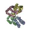


 PDBj
PDBj Assembly
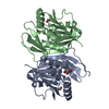

 Mass: 92.094 Da / Num. of mol.: 3 / Source method: obtained synthetically / Formula: C3H8O3
 Mass: 18.015 Da / Num. of mol.: 233 / Source method: isolated from a natural source / Formula: H2O
 Sample preparation
 / Beamline: X29A / Wavelength: 0.9791
  Processing