 Movie
Movie Controller

[English] 日本語
 Yorodumi
- PDB-3ghe: Crystal structure of anti-HIV-1 Fab 537-10D in complex with V3 pe... -
+ Open data

- Basic information
| Entry | Database: PDB / ID: 3ghe | ||||||
|---|---|---|---|---|---|---|---|
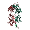
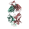
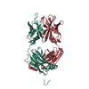
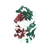
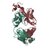
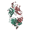
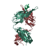
| Title | Crystal structure of anti-HIV-1 Fab 537-10D in complex with V3 peptide MN | ||||||
 Components Components |
| ||||||
 Keywords Keywords | IMMUNE SYSTEM / HIV / V3 LOOP / ANTIBODY-ANTIGEN INTERACTIONS / Envelope protein | ||||||
| Function / homology |  Function and homology information Function and homology informationmembrane fusion involved in viral entry into host cell / viral envelope / symbiont entry into host cell / virion attachment to host cell / virion membrane Similarity search - Function | ||||||
| Biological species |  Homo sapiens (human) Homo sapiens (human)  Human immunodeficiency virus 1 Human immunodeficiency virus 1 | ||||||
| Method |  X-RAY DIFFRACTION / SYNCHROTRON / MOLECULAR REPLACEMENT / Resolution: 2.4 Å X-RAY DIFFRACTION / SYNCHROTRON / MOLECULAR REPLACEMENT / Resolution: 2.4 Å | ||||||
 Authors Authors | Kong, X.P. / Burke, V.J. | ||||||
 Citation Citation | Journal: Structure / Year: 2009 Title: Structural basis of the cross-reactivity of genetically related human anti-HIV-1 mAbs: implications for design of V3-based immunogens Authors: Burke, V. / Williams, C. / Sukumaran, M. / Kim, S.S. / Li, H. / Wang, X.H. / Gorny, M.K. / Zolla-Pazner, S. / Kong, X.P. | ||||||
| History |
|
- Structure visualization
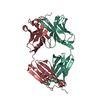
| Structure viewer | Molecule: MolmilJmol/JSmol |
|---|
- Downloads & links
-Download
| PDBx/mmCIF format | 3ghe.cif.gz | 106.6 KB | Display | PDBx/mmCIF format |
|---|---|---|---|---|
| PDB format | pdb3ghe.ent.gz | 79.4 KB | Display | PDB format |
| PDBx/mmJSON format | 3ghe.json.gz | Tree view | PDBx/mmJSON format | |
| Others |  Other downloads Other downloads |
-Validation report
| Summary document | 3ghe_validation.pdf.gz | 426.5 KB | Display | wwPDB validaton report |
|---|---|---|---|---|
| Full document | 3ghe_full_validation.pdf.gz | 431.9 KB | Display | |
| Data in XML | 3ghe_validation.xml.gz | 21.6 KB | Display | |
| Data in CIF | 3ghe_validation.cif.gz | 31.3 KB | Display | |
| Arichive directory | https://data.pdbj.org/pub/pdb/validation_reports/gh/3gheftp://data.pdbj.org/pub/pdb/validation_reports/gh/3ghe | HTTPS FTP |
-Related structure data


-Links
 PDBj
PDBj


- Assembly
| Deposited unit | 
| ||||||||
|---|---|---|---|---|---|---|---|---|---|
| 1 |
| ||||||||
| Unit cell |
|
-Components
| #1: Antibody | Mass: 22911.236 Da / Num. of mol.: 1 / Source method: isolated from a natural source / Details: B cell / Source: (natural) Homo sapiens (human) |
|---|---|
| #2: Antibody | Mass: 25433.422 Da / Num. of mol.: 1 / Source method: isolated from a natural source / Details: B cell / Source: (natural) Homo sapiens (human) |
| #3: Protein/peptide | Mass: 1748.064 Da / Num. of mol.: 1 / Source method: obtained synthetically / Source: (synth.) Human immunodeficiency virus 1 / References: UniProt: P88403 |
| #4: Water | ChemComp-HOH /  Mass: 18.015 Da / Num. of mol.: 322 / Source method: isolated from a natural source / Formula: H2O Mass: 18.015 Da / Num. of mol.: 322 / Source method: isolated from a natural source / Formula: H2O |
| Has protein modification | Y |
-Experimental details
-Experiment
| Experiment | Method: X-RAY DIFFRACTION / Number of used crystals: 1 |
|---|
- Sample preparation
| Crystal | Density Matthews: 2.47 Å3/Da / Density % sol: 50.15 % |
|---|---|
| Crystal grow | Temperature: 296.15 K / pH: 7.5 Details: 20% PEG 4000, 5% Isoproponal, 0.1M Hepes pH 7.5, VAPOR DIFFUSION, HANGING DROP, temperature 296.15K |
-Data collection
| Diffraction | Mean temperature: 100 K |
|---|---|
| Diffraction source | Source: SYNCHROTRON / Site: NSLS  / Beamline: X4C / Wavelength: 0.979 / Beamline: X4C / Wavelength: 0.979 |
| Detector | Type: MAR CCD 165 mm / Detector: CCD / Date: Feb 29, 2008 / Details: MIRRORS |
| Radiation | Monochromator: SI(111) / Protocol: SINGLE WAVELENGTH / Monochromatic (M) / Laue (L): M / Scattering type: x-ray |
| Radiation wavelength | Wavelength: 0.979 Å / Relative weight: 1 |
| Reflection | Resolution: 1.99→29.95 Å / Num. obs: 23276 / % possible obs: 73.1 % / Observed criterion σ(I): 0 / Redundancy: 5.4 % / Biso Wilson estimate: 31.8 Å2 / Rmerge(I) obs: 0.085 / Rsym value: 0.08 |
| Reflection shell | Resolution: 1.99→2.06 Å / Redundancy: 1.4 % / Rmerge(I) obs: 0.494 / Mean I/σ(I) obs: 2 / Rsym value: 0.331 / % possible all: 8 |
- Processing
| Software |
| ||||||||||||||||||||||||||||||||||||||||||||||||||||||||||||
|---|---|---|---|---|---|---|---|---|---|---|---|---|---|---|---|---|---|---|---|---|---|---|---|---|---|---|---|---|---|---|---|---|---|---|---|---|---|---|---|---|---|---|---|---|---|---|---|---|---|---|---|---|---|---|---|---|---|---|---|---|---|
| Refinement | Method to determine structure: MOLECULAR REPLACEMENT Starting model: CRYSTAL STRUCTURE OF 2557 FAB WITH V3 PEPTIDE NY5 Resolution: 2.4→29.95 Å / Occupancy max: 1 / Occupancy min: 1 / Isotropic thermal model: RESTRAINED / Cross valid method: THROUGHOUT / σ(F): 0 / Stereochemistry target values: ENGH & HUBER
| ||||||||||||||||||||||||||||||||||||||||||||||||||||||||||||
| Solvent computation | Bsol: 43.64 Å2 | ||||||||||||||||||||||||||||||||||||||||||||||||||||||||||||
| Displacement parameters | Biso mean: 34.58 Å2
| ||||||||||||||||||||||||||||||||||||||||||||||||||||||||||||
| Refine analyze |
| ||||||||||||||||||||||||||||||||||||||||||||||||||||||||||||
| Refinement step | Cycle: LAST / Resolution: 2.4→29.95 Å
| ||||||||||||||||||||||||||||||||||||||||||||||||||||||||||||
| Refine LS restraints |
| ||||||||||||||||||||||||||||||||||||||||||||||||||||||||||||
| LS refinement shell | Resolution: 2.4→2.55 Å / Rfactor Rfree error: 0.03
| ||||||||||||||||||||||||||||||||||||||||||||||||||||||||||||
| Xplor file |
|
