 Movie
Movie Controller

[English] 日本語
 Yorodumi
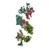
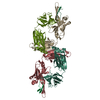
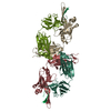
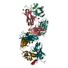
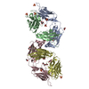
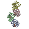
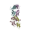
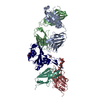
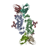
- PDB-1q1j: Crystal Structure Analysis of anti-HIV-1 Fab 447-52D in complex w... -
+ Open data

- Basic information
| Entry | Database: PDB / ID: 1q1j | ||||||
|---|---|---|---|---|---|---|---|
| Title | Crystal Structure Analysis of anti-HIV-1 Fab 447-52D in complex with V3 peptide | ||||||
 Components Components |
| ||||||
 Keywords Keywords | IMMUNE SYSTEM / Fab-peptide complex / HIV-1 / gp120 / V3 loop | ||||||
| Function / homology |  Function and homology information Function and homology informationDectin-2 family / symbiont-mediated perturbation of host defense response / positive regulation of plasma membrane raft polarization / positive regulation of receptor clustering / host cell endosome membrane / clathrin-dependent endocytosis of virus by host cell / viral protein processing / fusion of virus membrane with host plasma membrane / fusion of virus membrane with host endosome membrane / viral envelope ...Dectin-2 family / symbiont-mediated perturbation of host defense response / positive regulation of plasma membrane raft polarization / positive regulation of receptor clustering / host cell endosome membrane / clathrin-dependent endocytosis of virus by host cell / viral protein processing / fusion of virus membrane with host plasma membrane / fusion of virus membrane with host endosome membrane / viral envelope / virion attachment to host cell / host cell plasma membrane / virion membrane / structural molecule activity / membrane / identical protein binding Similarity search - Function | ||||||
| Biological species |  Homo sapiens (human) Homo sapiens (human) | ||||||
| Method |  X-RAY DIFFRACTION / SYNCHROTRON / MOLECULAR REPLACEMENT / Resolution: 2.5 Å X-RAY DIFFRACTION / SYNCHROTRON / MOLECULAR REPLACEMENT / Resolution: 2.5 Å | ||||||
 Authors Authors | Stanfield, R.L. / Gorny, M.K. / Williams, C. / Zolla-Pazner, S. / Wilson, I.A. | ||||||
 Citation Citation | Journal: Structure / Year: 2004 Title: Structural rationale for the broad neutralization of HIV-1 by human monoclonal antibody 447-52D. Authors: Stanfield, R.L. / Gorny, M.K. / Williams, C. / Zolla-Pazner, S. / Wilson, I.A. | ||||||
| History |
|
- Structure visualization
| Structure viewer | Molecule: MolmilJmol/JSmol |
|---|
- Downloads & links
-Download
| PDBx/mmCIF format | 1q1j.cif.gz | 177.6 KB | Display | PDBx/mmCIF format |
|---|---|---|---|---|
| PDB format | pdb1q1j.ent.gz | 141.5 KB | Display | PDB format |
| PDBx/mmJSON format | 1q1j.json.gz | Tree view | PDBx/mmJSON format | |
| Others |  Other downloads Other downloads |
-Validation report
| Arichive directory | https://data.pdbj.org/pub/pdb/validation_reports/q1/1q1jftp://data.pdbj.org/pub/pdb/validation_reports/q1/1q1j | HTTPS FTP |
|---|
-Related structure data
| Related structure data | 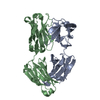 8fabS S: Starting model for refinement |
|---|---|
| Similar structure data |

-Links
 PDBj
PDBj


- Assembly
| Deposited unit | 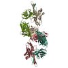
| ||||||||||
|---|---|---|---|---|---|---|---|---|---|---|---|
| 1 |
| ||||||||||
| Unit cell |
|
-Components
| #1: Antibody | Mass: 22684.111 Da / Num. of mol.: 2 / Source method: isolated from a natural source / Details: isolated from peripheral blood cells / Source: (natural) Homo sapiens (human) / Organ: blood#2: Antibody | Mass: 24763.775 Da / Num. of mol.: 2 / Source method: isolated from a natural source / Details: isolated from peripheral blood cells / Source: (natural) Homo sapiens (human) / Organ: blood#3: Protein/peptide | Mass: 1827.182 Da / Num. of mol.: 2 / Source method: obtained synthetically / Details: This sequence occurs in HIV-1 gp120 / References: UniProt: P05877*PLUS #4: Water | ChemComp-HOH / |  Mass: 18.015 Da / Num. of mol.: 53 / Source method: isolated from a natural source / Formula: H2O Mass: 18.015 Da / Num. of mol.: 53 / Source method: isolated from a natural source / Formula: H2OHas protein modification | Y | |
|---|
-Experimental details
-Experiment
| Experiment | Method: X-RAY DIFFRACTION / Number of used crystals: 1 |
|---|
- Sample preparation
| Crystal | Density Matthews: 2.93 Å3/Da / Density % sol: 57.7 % | ||||||||||||||||||||||||||||||
|---|---|---|---|---|---|---|---|---|---|---|---|---|---|---|---|---|---|---|---|---|---|---|---|---|---|---|---|---|---|---|---|
| Crystal grow | Temperature: 298 K / Method: vapor diffusion, sitting drop / pH: 6 Details: 1.23M ammonium sulfate, 0.1M sodium cacodylate, pH 6.0, VAPOR DIFFUSION, SITTING DROP, temperature 298.0K | ||||||||||||||||||||||||||||||
| Crystal grow | *PLUS Method: vapor diffusion, sitting drop | ||||||||||||||||||||||||||||||
| Components of the solutions | *PLUS
|
-Data collection
| Diffraction | Mean temperature: 100 K |
|---|---|
| Diffraction source | Source: SYNCHROTRON / Site: SSRL  / Beamline: BL11-1 / Wavelength: 0.984 Å / Beamline: BL11-1 / Wavelength: 0.984 Å |
| Detector | Type: ADSC QUANTUM 315 / Detector: CCD / Date: Jul 17, 2002 |
| Radiation | Protocol: SINGLE WAVELENGTH / Monochromatic (M) / Laue (L): M / Scattering type: x-ray |
| Radiation wavelength | Wavelength: 0.984 Å / Relative weight: 1 |
| Reflection | Resolution: 2.5→29.8 Å / Num. all: 35179 / Num. obs: 35179 / % possible obs: 90.7 % / Observed criterion σ(F): 0 / Observed criterion σ(I): 0 / Redundancy: 1.9 % / Biso Wilson estimate: 49.9 Å2 / Rsym value: 0.077 / Net I/σ(I): 13.7 |
| Reflection shell | Resolution: 2.5→2.54 Å / Mean I/σ(I) obs: 1.9 / Rsym value: 0.531 / % possible all: 0.952 |
| Reflection | *PLUS Lowest resolution: 30 Å / Num. measured all: 67571 / Rmerge(I) obs: 0.077 |
| Reflection shell | *PLUS % possible obs: 95.2 % / Num. unique obs: 1816 / Num. measured obs: 3553 / Rmerge(I) obs: 0.531 |
- Processing
| Software |
| |||||||||||||||||||||||||
|---|---|---|---|---|---|---|---|---|---|---|---|---|---|---|---|---|---|---|---|---|---|---|---|---|---|---|
| Refinement | Method to determine structure: MOLECULAR REPLACEMENT Starting model: PDB ENTRY 8FAB Resolution: 2.5→29.8 Å / Rfactor Rfree error: 0.007 / Isotropic thermal model: RESTRAINED / Cross valid method: THROUGHOUT / σ(F): 0 / σ(I): 0 / Stereochemistry target values: Engh & Huber
| |||||||||||||||||||||||||
| Solvent computation | Solvent model: FLAT MODEL / Bsol: 26.3505 Å2 / ksol: 0.337844 e/Å3 | |||||||||||||||||||||||||
| Displacement parameters | Biso mean: 48.1 Å2
| |||||||||||||||||||||||||
| Refine analyze |
| |||||||||||||||||||||||||
| Refinement step | Cycle: LAST / Resolution: 2.5→29.8 Å
| |||||||||||||||||||||||||
| Refine LS restraints |
| |||||||||||||||||||||||||
| LS refinement shell | Resolution: 2.5→2.59 Å / Total num. of bins used: 6
| |||||||||||||||||||||||||
| Xplor file |
| |||||||||||||||||||||||||
| Refinement | *PLUS Lowest resolution: 30 Å / % reflection Rfree: 5 % / Rfactor Rwork: 0.249 | |||||||||||||||||||||||||
| Solvent computation | *PLUS | |||||||||||||||||||||||||
| Displacement parameters | *PLUS | |||||||||||||||||||||||||
| Refine LS restraints | *PLUS
| |||||||||||||||||||||||||
| LS refinement shell | *PLUS Rfactor Rfree: 0.51 |
