 Movie
Movie Controller

[English] 日本語
 Yorodumi
- PDB-3ggz: Crystal Structure of S.cerevisiae Ist1 N-terminal domain in compl... -
+ Open data

- Basic information
| Entry | Database: PDB / ID: 3ggz | ||||||
|---|---|---|---|---|---|---|---|
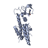
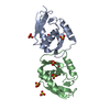
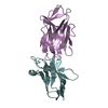
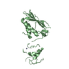
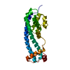
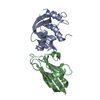
| Title | Crystal Structure of S.cerevisiae Ist1 N-terminal domain in complex with Did2 MIM motif | ||||||
 Components Components |
| ||||||
 Keywords Keywords | PROTEIN TRANSPORT / ENDOCYTOSIS / novel MIM binding mode / Phosphoprotein / Coiled coil / Endosome / Membrane / Transport | ||||||
| Function / homology |  Function and homology information Function and homology informationESCRT III complex assembly / Sealing of the nuclear envelope (NE) by ESCRT-III / ESCRT III complex / endosome transport via multivesicular body sorting pathway / late endosome to vacuole transport via multivesicular body sorting pathway / endosome to plasma membrane protein transport / protein targeting to vacuole / ATPase inhibitor activity / late endosome to vacuole transport / Neutrophil degranulation ...ESCRT III complex assembly / Sealing of the nuclear envelope (NE) by ESCRT-III / ESCRT III complex / endosome transport via multivesicular body sorting pathway / late endosome to vacuole transport via multivesicular body sorting pathway / endosome to plasma membrane protein transport / protein targeting to vacuole / ATPase inhibitor activity / late endosome to vacuole transport / Neutrophil degranulation / multivesicular body / intracellular protein localization / late endosome / protein transport / endosome / cytoplasm Similarity search - Function | ||||||
| Biological species |  | ||||||
| Method |  X-RAY DIFFRACTION / SYNCHROTRON / MOLECULAR REPLACEMENT / molecular replacement / Resolution: 3.8 Å X-RAY DIFFRACTION / SYNCHROTRON / MOLECULAR REPLACEMENT / molecular replacement / Resolution: 3.8 Å | ||||||
 Authors Authors | Xiao, J. / Xu, Z. | ||||||
 Citation Citation | Journal: MOLECULAR BIOLOGY OF THE CELL / Year: 2009 Title: Structural basis of Ist1 function and Ist1-Did2 interaction in the multivesicular body pathway and cytokinesis. Authors: Xiao, J. / Chen, X.W. / Davies, B.A. / Saltiel, A.R. / Katzmann, D.J. / Xu, Z. | ||||||
| History |
|
- Structure visualization
| Structure viewer | Molecule: MolmilJmol/JSmol |
|---|
- Downloads & links
-Download
| PDBx/mmCIF format | 3ggz.cif.gz | 169.1 KB | Display | PDBx/mmCIF format |
|---|---|---|---|---|
| PDB format | pdb3ggz.ent.gz | 137 KB | Display | PDB format |
| PDBx/mmJSON format | 3ggz.json.gz | Tree view | PDBx/mmJSON format | |
| Others |  Other downloads Other downloads |
-Validation report
| Arichive directory | https://data.pdbj.org/pub/pdb/validation_reports/gg/3ggzftp://data.pdbj.org/pub/pdb/validation_reports/gg/3ggz | HTTPS FTP |
|---|
-Related structure data


-Links
 PDBj
PDBj


- Assembly
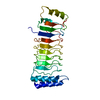
| Deposited unit | 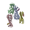
| |||||||||||||||||||||||||||||||||||
|---|---|---|---|---|---|---|---|---|---|---|---|---|---|---|---|---|---|---|---|---|---|---|---|---|---|---|---|---|---|---|---|---|---|---|---|---|
| 1 | 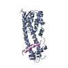
| |||||||||||||||||||||||||||||||||||
| 2 | 
| |||||||||||||||||||||||||||||||||||
| 3 | 
| |||||||||||||||||||||||||||||||||||
| 4 | 
| |||||||||||||||||||||||||||||||||||
| Unit cell |
| |||||||||||||||||||||||||||||||||||
| Noncrystallographic symmetry (NCS) | NCS domain:
NCS domain segments: Dom-ID: 1 / Component-ID: 1 / Beg auth comp-ID: PHE / Beg label comp-ID: PHE / End auth comp-ID: TYR / End label comp-ID: TYR / Auth seq-ID: 8 - 180 / Label seq-ID: 8 - 180
NCS ensembles :
|
-Components
| #1: Protein | Mass: 22380.164 Da / Num. of mol.: 4 / Fragment: N-terminal domain, UNP residues 1-192 Source method: isolated from a genetically manipulated source Source: (gene. exp.) Gene: IST1, N0809, YNL265C / Production host:  #2: Protein/peptide | Mass: 3341.793 Da / Num. of mol.: 4 / Fragment: MIM motif, UNP residues 176-204 Source method: isolated from a genetically manipulated source Source: (gene. exp.) Gene: CHM1, DID2, FTI1, VPS46, YKR035W-A / Production host: |
|---|
-Experimental details
-Experiment
| Experiment | Method: X-RAY DIFFRACTION / Number of used crystals: 1 |
|---|
- Sample preparation
| Crystal | Density Matthews: 4.08 Å3/Da / Density % sol: 69.85 % |
|---|---|
| Crystal grow | Temperature: 293 K / Method: vapor diffusion / Details: Sodium Citrate, vapor diffusion, temperature 293K |
-Data collection
| Diffraction | Mean temperature: 100 K | ||||||||||||||||||||||||||||||||||||||||||||||||||||||||||||||||||||||||||||||||||||
|---|---|---|---|---|---|---|---|---|---|---|---|---|---|---|---|---|---|---|---|---|---|---|---|---|---|---|---|---|---|---|---|---|---|---|---|---|---|---|---|---|---|---|---|---|---|---|---|---|---|---|---|---|---|---|---|---|---|---|---|---|---|---|---|---|---|---|---|---|---|---|---|---|---|---|---|---|---|---|---|---|---|---|---|---|---|
| Diffraction source | Source: SYNCHROTRON / Site: APS  / Beamline: 21-ID-D / Wavelength: 0.9793 Å / Beamline: 21-ID-D / Wavelength: 0.9793 Å | ||||||||||||||||||||||||||||||||||||||||||||||||||||||||||||||||||||||||||||||||||||
| Detector | Detector: CCD / Date: Apr 1, 2008 | ||||||||||||||||||||||||||||||||||||||||||||||||||||||||||||||||||||||||||||||||||||
| Radiation | Protocol: SINGLE WAVELENGTH / Monochromatic (M) / Laue (L): M / Scattering type: x-ray | ||||||||||||||||||||||||||||||||||||||||||||||||||||||||||||||||||||||||||||||||||||
| Radiation wavelength | Wavelength: 0.9793 Å / Relative weight: 1 | ||||||||||||||||||||||||||||||||||||||||||||||||||||||||||||||||||||||||||||||||||||
| Reflection | Resolution: 3.8→50 Å / Num. obs: 17219 / % possible obs: 99.6 % / Redundancy: 6 % / Rmerge(I) obs: 0.091 / Net I/σ(I): 16.617 | ||||||||||||||||||||||||||||||||||||||||||||||||||||||||||||||||||||||||||||||||||||
| Reflection shell |
|
-Phasing
| Phasing | Method: molecular replacement | |||||||||
|---|---|---|---|---|---|---|---|---|---|---|
| Phasing MR | Rfactor: 49.32 / Model details: Phaser MODE: MR_AUTO
|
- Processing
| Software |
| ||||||||||||||||||||||||||||||||||||||||||||||||||||||||||||||||||||||||||||||||
|---|---|---|---|---|---|---|---|---|---|---|---|---|---|---|---|---|---|---|---|---|---|---|---|---|---|---|---|---|---|---|---|---|---|---|---|---|---|---|---|---|---|---|---|---|---|---|---|---|---|---|---|---|---|---|---|---|---|---|---|---|---|---|---|---|---|---|---|---|---|---|---|---|---|---|---|---|---|---|---|---|---|
| Refinement | Method to determine structure: MOLECULAR REPLACEMENT / Resolution: 3.8→49.03 Å / Rfactor Rfree error: 0.01 / Occupancy max: 1 / Occupancy min: 1 / Data cutoff high absF: 5432034.24 / Data cutoff low absF: 0 / Isotropic thermal model: RESTRAINED / Cross valid method: THROUGHOUT / σ(F): 0
| ||||||||||||||||||||||||||||||||||||||||||||||||||||||||||||||||||||||||||||||||
| Solvent computation | Solvent model: FLAT MODEL / Bsol: 104.977 Å2 / ksol: 0.35 e/Å3 | ||||||||||||||||||||||||||||||||||||||||||||||||||||||||||||||||||||||||||||||||
| Displacement parameters | Biso mean: 146.1 Å2
| ||||||||||||||||||||||||||||||||||||||||||||||||||||||||||||||||||||||||||||||||
| Refine analyze |
| ||||||||||||||||||||||||||||||||||||||||||||||||||||||||||||||||||||||||||||||||
| Refinement step | Cycle: LAST / Resolution: 3.8→49.03 Å
| ||||||||||||||||||||||||||||||||||||||||||||||||||||||||||||||||||||||||||||||||
| Refine LS restraints |
| ||||||||||||||||||||||||||||||||||||||||||||||||||||||||||||||||||||||||||||||||
| Refine LS restraints NCS | NCS model details: CONSTR | ||||||||||||||||||||||||||||||||||||||||||||||||||||||||||||||||||||||||||||||||
| LS refinement shell | Resolution: 3.8→4.04 Å / Rfactor Rfree error: 0.03 / Total num. of bins used: 6
| ||||||||||||||||||||||||||||||||||||||||||||||||||||||||||||||||||||||||||||||||
| Xplor file |
|
