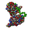
 Movie
Movie Controller

[English] 日本語
 Yorodumi
- PDB-3gfk: Crystal structure of Bacillus subtilis Spx/RNA polymerase alpha s... -
+ Open data

- Basic information
| Entry | Database: PDB / ID: 3gfk | ||||||
|---|---|---|---|---|---|---|---|
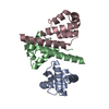
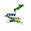
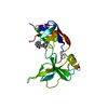
| Title | Crystal structure of Bacillus subtilis Spx/RNA polymerase alpha subunit C-terminal domain complex | ||||||
 Components Components |
| ||||||
 Keywords Keywords | Transcription/Transferase / protein-protein complex / Cytoplasm / Redox-active center / Stress response / Transcription / Transcription regulation / DNA-directed RNA polymerase / Nucleotidyltransferase / Transferase / Transcription-Transferase COMPLEX | ||||||
| Function / homology |  Function and homology information Function and homology informationDNA-directed RNA polymerase complex / DNA-directed RNA polymerase / DNA-directed RNA polymerase activity / protein dimerization activity / negative regulation of DNA-templated transcription / DNA-templated transcription / DNA binding / cytoplasm Similarity search - Function | ||||||
| Biological species |  | ||||||
| Method |  X-RAY DIFFRACTION / SYNCHROTRON / MOLECULAR REPLACEMENT / Resolution: 2.3 Å X-RAY DIFFRACTION / SYNCHROTRON / MOLECULAR REPLACEMENT / Resolution: 2.3 Å | ||||||
 Authors Authors | Lamour, V. / Westblade, L.F. / Campbell, E.A. / Darst, S.A. | ||||||
 Citation Citation | Journal: J.Struct.Biol. / Year: 2009 Title: Crystal structure of the in vivo-assembled Bacillus subtilis Spx/RNA polymerase alpha subunit C-terminal domain complex Authors: Lamour, V. / Westblade, L.F. / Campbell, E.A. / Darst, S.A. | ||||||
| History |
|
- Structure visualization
| Structure viewer | Molecule: MolmilJmol/JSmol |
|---|
- Downloads & links
-Download
| PDBx/mmCIF format | 3gfk.cif.gz | 58.4 KB | Display | PDBx/mmCIF format |
|---|---|---|---|---|
| PDB format | pdb3gfk.ent.gz | 41.6 KB | Display | PDB format |
| PDBx/mmJSON format | 3gfk.json.gz | Tree view | PDBx/mmJSON format | |
| Others |  Other downloads Other downloads |
-Validation report
| Summary document | 3gfk_validation.pdf.gz | 408.4 KB | Display | wwPDB validaton report |
|---|---|---|---|---|
| Full document | 3gfk_full_validation.pdf.gz | 416.6 KB | Display | |
| Data in XML | 3gfk_validation.xml.gz | 7.1 KB | Display | |
| Data in CIF | 3gfk_validation.cif.gz | 10.7 KB | Display | |
| Arichive directory | https://data.pdbj.org/pub/pdb/validation_reports/gf/3gfkftp://data.pdbj.org/pub/pdb/validation_reports/gf/3gfk | HTTPS FTP |
-Related structure data
| Related structure data | 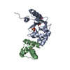 1z3eS S: Starting model for refinement |
|---|---|
| Similar structure data |


-Links
 PDBj
PDBj

- Assembly
| Deposited unit | 
| ||||||||
|---|---|---|---|---|---|---|---|---|---|
| 1 |
| ||||||||
| Unit cell |
|
-Components
| #1: Protein | Mass: 15551.892 Da / Num. of mol.: 1 Source method: isolated from a genetically manipulated source Source: (gene. exp.) |
|---|---|
| #2: Protein | Mass: 9118.472 Da / Num. of mol.: 1 / Fragment: Alpha C-terminal domain (alpha-CTD) Source method: isolated from a genetically manipulated source Source: (gene. exp.) |
| #3: Water | ChemComp-HOH /  Mass: 18.015 Da / Num. of mol.: 257 / Source method: isolated from a natural source / Formula: H2O Mass: 18.015 Da / Num. of mol.: 257 / Source method: isolated from a natural source / Formula: H2O |
| Has protein modification | Y |
-Experimental details
-Experiment
| Experiment | Method: X-RAY DIFFRACTION / Number of used crystals: 1 |
|---|
- Sample preparation
| Crystal | Density Matthews: 2.2 Å3/Da / Density % sol: 44.2 % |
|---|---|
| Crystal grow | Temperature: 295 K / Method: vapor diffusion / pH: 5 Details: vapor diffusion against 6% (w/v) PEG 6000, 100 mM sodium acetate, pH 5.0, 10 micromolar CuCl2, protein concentration 11.5 mg/mL, temperature 295K |
-Data collection
| Diffraction | Mean temperature: 180 K |
|---|---|
| Diffraction source | Source: SYNCHROTRON / Site: NSLS  / Beamline: X25 / Wavelength: 1.3376 / Beamline: X25 / Wavelength: 1.3376 |
| Detector | Type: ADSC QUANTUM 315 / Detector: CCD / Date: Jan 20, 2004 |
| Radiation | Protocol: SINGLE WAVELENGTH / Monochromatic (M) / Laue (L): M / Scattering type: x-ray |
| Radiation wavelength | Wavelength: 1.3376 Å / Relative weight: 1 |
| Reflection | Resolution: 2.2→20 Å / Num. obs: 11313 / % possible obs: 99.4 % |
- Processing
| Software |
| |||||||||||||||||||||||||
|---|---|---|---|---|---|---|---|---|---|---|---|---|---|---|---|---|---|---|---|---|---|---|---|---|---|---|
| Refinement | Method to determine structure: MOLECULAR REPLACEMENT Starting model: PDB ENTRY 1Z3E Resolution: 2.3→20 Å / Isotropic thermal model: ANISOTROPIC / Cross valid method: THROUGHOUT / σ(F): 0 / Stereochemistry target values: ENGH & HUBER
| |||||||||||||||||||||||||
| Displacement parameters |
| |||||||||||||||||||||||||
| Refinement step | Cycle: LAST / Resolution: 2.3→20 Å
| |||||||||||||||||||||||||
| Xplor file |
|
