- PDB-3gf6: Crystal structure of a bacterial lipoprotein (bt_1233) from bacte... -
+
Open data
ID or keywords:
Loading...
-
Basic information
Entry
Database: PDB / ID: 3gf6
Title
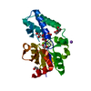
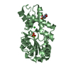
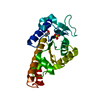
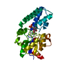
Crystal structure of a bacterial lipoprotein (bt_1233) from bacteroides thetaiotaomicron vpi-5482 at 1.69 A resolution
Components
uncharacterized bacterial lipoprotein
Keywords
UNKNOWN FUNCTION / All-beta fold / structural genomics / Joint Center for Structural Genomics / JCSG / Protein Structure Initiative / PSI-2
Function / homology
Protein of unknown function DUF3845 / Protein of unknown function DUF3845 / Domain of Unknown Function with PDB structure / Jelly Rolls / Sandwich / Mainly Beta / DUF3845 domain-containing protein
Function and homology information
Biological species
Bacteroides thetaiotaomicron VPI-5482 (bacteria)
Method
X-RAY DIFFRACTION / SYNCHROTRON / SAD / Resolution: 1.69 Å
Mass: 18.015 Da / Num. of mol.: 664 / Source method: isolated from a natural source / Formula: H2O
Has protein modification
Y
Sequence details
THE CONSTRUCT (RESIDUES 22-244) WAS EXPRESSED WITH A PURIFICATION TAG MGSDKIHHHHHHENLYFQG. THE TAG ...THE CONSTRUCT (RESIDUES 22-244) WAS EXPRESSED WITH A PURIFICATION TAG MGSDKIHHHHHHENLYFQG. THE TAG WAS REMOVED WITH TEV PROTEASE LEAVING ONLY A GLYCINE (0) FOLLOWED BY THE TARGET SEQUENCE.
-
Experimental details
-
Experiment
Experiment
Method: X-RAY DIFFRACTION / Number of used crystals: 1
-
Sample preparation
Crystal
Density Matthews: 2.18 Å3/Da / Density % sol: 43.62 % Description: THE AND R MERGE VALUES REPORTED HERE ARE BASED ON TREATING FRIEDEL PAIRS AS SEPARATE REFLECTIONS.
Crystal grow
Temperature: 277 K / Method: vapor diffusion, sitting drop / pH: 6.5 Details: 31.3000% polyethylene glycol 6000, 0.1M MES pH 6.5, NANODROP, VAPOR DIFFUSION, SITTING DROP, temperature 277K
Monochromator: Double crystal monochromator / Protocol: SINGLE WAVELENGTH / Monochromatic (M) / Laue (L): M / Scattering type: x-ray
Radiation wavelength
Wavelength: 0.97951 Å / Relative weight: 1
Reflection
Resolution: 1.69→48.337 Å / Num. obs: 51502 / % possible obs: 99.9 % / Observed criterion σ(I): -3 / Biso Wilson estimate: 17.776 Å2 / Rmerge(I) obs: 0.105 / Net I/σ(I): 11.7
Reflection shell
Resolution (Å)
Rmerge(I) obs
Mean I/σ(I) obs
Num. measured obs
Num. unique obs
Diffraction-ID
% possible all
1.69-1.75
0.637
1.9
18320
5064
1
100
1.75-1.82
0.507
2.4
18376
5050
1
100
1.82-1.9
0.366
3.3
18048
4962
1
99.9
1.9-2
0.249
4.8
18470
5076
1
99.9
2-2.13
0.275
6.7
26598
5306
1
99.9
2.13-2.29
0.251
8.8
31144
4989
1
99.9
2.29-2.52
0.215
11.3
37751
5175
1
100
2.52-2.89
0.138
15.7
38157
5237
1
100
2.89-3.63
0.069
25.2
37458
5169
1
100
3.63-48.337
0.046
34.7
37878
5473
1
99.5
-
Phasing
Phasing
Method: SAD
-
Processing
Software
Name
Version
Classification
NB
REFMAC
5.5.0053
refinement
PHENIX
refinement
SHELX
phasing
MolProbity
3beta29
modelbuilding
XSCALE
datascaling
PDB_EXTRACT
3.006
dataextraction
XDS
datareduction
SHELXD
phasing
autoSHARP
phasing
Refinement
Method to determine structure: SAD / Resolution: 1.69→48.337 Å / Cor.coef. Fo:Fc: 0.966 / Cor.coef. Fo:Fc free: 0.939 / Occupancy max: 1 / Occupancy min: 0.09 / SU B: 5.153 / SU ML: 0.074 / TLS residual ADP flag: LIKELY RESIDUAL / Cross valid method: THROUGHOUT / σ(F): 0 / ESU R: 0.102 / ESU R Free: 0.108 Stereochemistry target values: MAXIMUM LIKELIHOOD WITH PHASES Details: 1. HYDROGENS HAVE BEEN ADDED IN THE RIDING POSITIONS. 2. A MET-INHIBITION PROTOCOL WAS USED FOR SELENOMETHIONINE INCORPORATION DURING PROTEIN EXPRESSION. THE OCCUPANCY OF THE SE ATOMS IN THE ...Details: 1. HYDROGENS HAVE BEEN ADDED IN THE RIDING POSITIONS. 2. A MET-INHIBITION PROTOCOL WAS USED FOR SELENOMETHIONINE INCORPORATION DURING PROTEIN EXPRESSION. THE OCCUPANCY OF THE SE ATOMS IN THE MSE RESIDUES WAS REDUCED TO 0.85 FOR THE REDUCED SCATTERING POWER DUE TO PARTIAL S-MET INCORPORATION. 3. ATOM RECORDS CONTAIN RESIDUAL B FACTORS ONLY. 4. ETHYLENE GLYCOLS (EDO) USED AS CRYOPROTECTANT WERE MODELED INTO THE STRUCTURE.
Rfactor
Num. reflection
% reflection
Selection details
Rfree
0.211
2615
5.1 %
RANDOM
Rwork
0.161
-
-
-
obs
0.163
51442
99.94 %
-
Solvent computation
Ion probe radii: 0.8 Å / Shrinkage radii: 0.8 Å / VDW probe radii: 1.2 Å / Solvent model: MASK
In the structure databanks used in Yorodumi, some data are registered as the other names, "COVID-19 virus" and "2019-nCoV". Here are the details of the virus and the list of structure data.
Jan 31, 2019. EMDB accession codes are about to change! (news from PDBe EMDB page)
EMDB accession codes are about to change! (news from PDBe EMDB page)
The allocation of 4 digits for EMDB accession codes will soon come to an end. Whilst these codes will remain in use, new EMDB accession codes will include an additional digit and will expand incrementally as the available range of codes is exhausted. The current 4-digit format prefixed with “EMD-” (i.e. EMD-XXXX) will advance to a 5-digit format (i.e. EMD-XXXXX), and so on. It is currently estimated that the 4-digit codes will be depleted around Spring 2019, at which point the 5-digit format will come into force.
The EM Navigator/Yorodumi systems omit the EMD- prefix.
Related info.:Q: What is EMD? / ID/Accession-code notation in Yorodumi/EM Navigator
Yorodumi is a browser for structure data from EMDB, PDB, SASBDB, etc.
This page is also the successor to EM Navigator detail page, and also detail information page/front-end page for Omokage search.
The word "yorodu" (or yorozu) is an old Japanese word meaning "ten thousand". "mi" (miru) is to see.
Related info.:EMDB / PDB / SASBDB / Comparison of 3 databanks / Yorodumi Search / Aug 31, 2016. New EM Navigator & Yorodumi / Yorodumi Papers / Jmol/JSmol / Function and homology information / Changes in new EM Navigator and Yorodumi
 Movie
Movie Controller

 Yorodumi
 Open data
 Basic information
 Components
 Keywords
 Function and homology information
 Bacteroides thetaiotaomicron VPI-5482 (bacteria)
 X-RAY DIFFRACTION /
  Authors
 Citation
 Structure visualization
 Downloads & links
 Other downloads
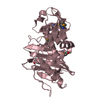


 PDBj
PDBj Assembly
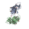

 Mass: 62.068 Da / Num. of mol.: 8 / Source method: obtained synthetically / Formula: C2H6O2
 Mass: 18.015 Da / Num. of mol.: 664 / Source method: isolated from a natural source / Formula: H2O
 Sample preparation
 / Beamline: BL9-2 / Wavelength: 0.97951
  Processing