 Movie
Movie Controller

[English] 日本語
 Yorodumi
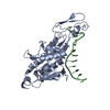
- PDB-3gf2: Crystal structure of the hypothetical regulator ST1710 complexed ... -
+ Open data

- Basic information
| Entry | Database: PDB / ID: 3gf2 | ||||||
|---|---|---|---|---|---|---|---|
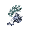
| Title | Crystal structure of the hypothetical regulator ST1710 complexed with sodium salicylate | ||||||
 Components Components | 146aa long hypothetical transcriptional regulator | ||||||
 Keywords Keywords | TRANSCRIPTION / Transcription regulator / ST1710 / MarR / DNA-binding / Transcription regulation / Structural Genomics / NPPSFA / National Project on Protein Structural and Functional Analyses / RIKEN Structural Genomics/Proteomics Initiative / RSGI | ||||||
| Function / homology |  Function and homology information Function and homology informationresponse to stress / DNA-binding transcription factor activity / metal ion binding Similarity search - Function | ||||||
| Biological species |   Sulfolobus tokodaii (archaea) Sulfolobus tokodaii (archaea) | ||||||
| Method |  X-RAY DIFFRACTION / Resolution: 1.8 Å X-RAY DIFFRACTION / Resolution: 1.8 Å | ||||||
 Authors Authors | Kumarevel, T. / Tanaka, T. / Yokoyama, S. / RIKEN Structural Genomics/Proteomics Initiative (RSGI) | ||||||
 Citation Citation | Journal: Nucleic Acids Res. / Year: 2009 Title: ST1710-DNA complex crystal structure reveals the DNA binding mechanism of the MarR family of regulators. Authors: Kumarevel, T. / Tanaka, T. / Umehara, T. / Yokoyama, S. #1: Journal: J.Struct.Biol. / Year: 2008Title: Crystal structure of the MarR family regulatory protein, ST1710, from Sulfolobus tokodaii strain 7. Authors: Kumarevel, T. / Tanaka, T. / Nishio, M. / Gopinath, S.C. / Takio, K. / Shinkai, A. / Kumar, P.K. / Yokoyama, S. | ||||||
| History |
|
- Structure visualization
| Structure viewer | Molecule: MolmilJmol/JSmol |
|---|
- Downloads & links
-Download
| PDBx/mmCIF format | 3gf2.cif.gz | 45.8 KB | Display | PDBx/mmCIF format |
|---|---|---|---|---|
| PDB format | pdb3gf2.ent.gz | 31.1 KB | Display | PDB format |
| PDBx/mmJSON format | 3gf2.json.gz | Tree view | PDBx/mmJSON format | |
| Others |  Other downloads Other downloads |
-Validation report
| Arichive directory | https://data.pdbj.org/pub/pdb/validation_reports/gf/3gf2ftp://data.pdbj.org/pub/pdb/validation_reports/gf/3gf2 | HTTPS FTP |
|---|
-Related structure data
| Related structure data |  3gezC 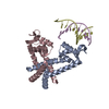 3gfiC  3gfjC  3gflC  3gfmC  2eb7S S: Starting model for refinement C: citing same article ( |
|---|---|
| Similar structure data | |
| Other databases |


-Links
 PDBj
PDBj

- Assembly
| Deposited unit | 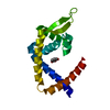
| ||||||||
|---|---|---|---|---|---|---|---|---|---|
| 1 | 
| ||||||||
| Unit cell |
|
-Components
| #1: Protein | Mass: 16879.564 Da / Num. of mol.: 1 Source method: isolated from a genetically manipulated source Source: (gene. exp.) Sulfolobus tokodaii (archaea) / Gene: ST1710 / Plasmid: pET21a / Production host:  |
|---|---|
| #2: Chemical | ChemComp-SAL / 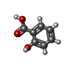  Mass: 138.121 Da / Num. of mol.: 1 / Source method: obtained synthetically / Formula: C7H6O3 Mass: 138.121 Da / Num. of mol.: 1 / Source method: obtained synthetically / Formula: C7H6O3 |
| #3: Water | ChemComp-HOH /  Mass: 18.015 Da / Num. of mol.: 152 / Source method: isolated from a natural source / Formula: H2O Mass: 18.015 Da / Num. of mol.: 152 / Source method: isolated from a natural source / Formula: H2O |
-Experimental details
-Experiment
| Experiment | Method: X-RAY DIFFRACTION / Number of used crystals: 1 |
|---|
- Sample preparation
| Crystal | Density Matthews: 2.17 Å3/Da / Density % sol: 43.3 % |
|---|---|
| Crystal grow | Temperature: 293 K / Method: vapor diffusion, sitting drop / pH: 6.5 Details: 18% PEG 8000, 0.2M Calcium acetate, 0.1M Sodium Cacodylate, pH 6.5, VAPOR DIFFUSION, SITTING DROP, temperature 293K |
-Data collection
| Diffraction | Mean temperature: 180 K |
|---|---|
| Diffraction source | Source: ROTATING ANODE / Type: RIGAKU FR-D / Wavelength: 1.54178 Å |
| Detector | Type: RIGAKU RAXIS VII / Detector: IMAGE PLATE / Date: Oct 14, 2008 |
| Radiation | Protocol: SINGLE WAVELENGTH / Monochromatic (M) / Laue (L): M / Scattering type: x-ray |
| Radiation wavelength | Wavelength: 1.54178 Å / Relative weight: 1 |
| Reflection | Resolution: 1.8→40 Å / Num. all: 13381 / Num. obs: 13381 / % possible obs: 91.3 % / Observed criterion σ(F): 2 / Observed criterion σ(I): 2 / Redundancy: 13.4 % / Biso Wilson estimate: 27.3 Å2 / Rsym value: 0.1 |
| Reflection shell | Resolution: 1.8→1.86 Å / Redundancy: 7.6 % / Num. unique all: 1425 / Rsym value: 0.264 / % possible all: 99.8 |
- Processing
| Software |
| ||||||||||||||||||||||||||||||||||||
|---|---|---|---|---|---|---|---|---|---|---|---|---|---|---|---|---|---|---|---|---|---|---|---|---|---|---|---|---|---|---|---|---|---|---|---|---|---|
| Refinement | Starting model: 2EB7 Resolution: 1.8→19.76 Å / Rfactor Rfree error: 0.009 / Occupancy max: 1 / Occupancy min: 1 / Data cutoff high absF: 271895 / Data cutoff low absF: 0 / Isotropic thermal model: RESTRAINED / Cross valid method: THROUGHOUT / σ(F): 0 / Stereochemistry target values: Engh & Huber
| ||||||||||||||||||||||||||||||||||||
| Solvent computation | Solvent model: FLAT MODEL / Bsol: 55.181 Å2 / ksol: 0.374 e/Å3 | ||||||||||||||||||||||||||||||||||||
| Displacement parameters | Biso max: 63.17 Å2 / Biso mean: 29.642 Å2 / Biso min: 10.78 Å2
| ||||||||||||||||||||||||||||||||||||
| Refine analyze |
| ||||||||||||||||||||||||||||||||||||
| Refinement step | Cycle: LAST / Resolution: 1.8→19.76 Å
| ||||||||||||||||||||||||||||||||||||
| Refine LS restraints |
| ||||||||||||||||||||||||||||||||||||
| LS refinement shell | Resolution: 1.8→1.91 Å / Rfactor Rfree error: 0.051 / Total num. of bins used: 6
| ||||||||||||||||||||||||||||||||||||
| Xplor file |
|
