 Movie
Movie Controller

[English] 日本語
 Yorodumi
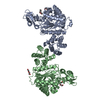
- PDB-1otp: STRUCTURAL AND THEORETICAL STUDIES SUGGEST DOMAIN MOVEMENT PRODUC... -
+ Open data

- Basic information
| Entry | Database: PDB / ID: 1otp | ||||||
|---|---|---|---|---|---|---|---|
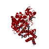
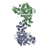
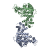
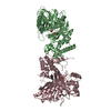
| Title | STRUCTURAL AND THEORETICAL STUDIES SUGGEST DOMAIN MOVEMENT PRODUCES AN ACTIVE CONFORMATION OF THYMIDINE PHOSPHORYLASE | ||||||
 Components Components | THYMIDINE PHOSPHORYLASE | ||||||
 Keywords Keywords | PHOSPHORYLASE / PYRIMIDINE METABOLISM / SALVAGE PATHWAY / DOMAIN MOVEMENT / TRANSFERASE / GLYCOSYLTRANSFERASE | ||||||
| Function / homology |  Function and homology information Function and homology informationthymidine phosphorylase / pyrimidine nucleoside metabolic process / thymidine phosphorylase activity / thymidine metabolic process / pyrimidine nucleobase metabolic process / 1,4-alpha-oligoglucan phosphorylase activity / DNA damage response / membrane / cytosol Similarity search - Function | ||||||
| Biological species |  | ||||||
| Method |  X-RAY DIFFRACTION / SINGLE ISOMORPHOUS REPLACEMENT / Resolution: 2.8 Å X-RAY DIFFRACTION / SINGLE ISOMORPHOUS REPLACEMENT / Resolution: 2.8 Å | ||||||
 Authors Authors | Pugmire, M.J. / Cook, W.J. / Jasanoff, A. / Walter, M.R. / Ealick, S.E. | ||||||
 Citation Citation | Journal: J.Mol.Biol. / Year: 1998 Title: Structural and theoretical studies suggest domain movement produces an active conformation of thymidine phosphorylase. Authors: Pugmire, M.J. / Cook, W.J. / Jasanoff, A. / Walter, M.R. / Ealick, S.E. | ||||||
| History |
|
- Structure visualization
| Structure viewer | Molecule: MolmilJmol/JSmol |
|---|
- Downloads & links
-Download
| PDBx/mmCIF format | 1otp.cif.gz | 90.3 KB | Display | PDBx/mmCIF format |
|---|---|---|---|---|
| PDB format | pdb1otp.ent.gz | 70 KB | Display | PDB format |
| PDBx/mmJSON format | 1otp.json.gz | Tree view | PDBx/mmJSON format | |
| Others |  Other downloads Other downloads |
-Validation report
| Arichive directory | https://data.pdbj.org/pub/pdb/validation_reports/ot/1otpftp://data.pdbj.org/pub/pdb/validation_reports/ot/1otp | HTTPS FTP |
|---|
-Related structure data


-Links
 PDBj
PDBj- Assembly
| Deposited unit | 
| ||||||||
|---|---|---|---|---|---|---|---|---|---|
| 1 | 
| ||||||||
| Unit cell |
|
-Components
| #1: Protein | Mass: 47240.988 Da / Num. of mol.: 1 / Source method: isolated from a natural source / Source: (natural) |
|---|
-Experimental details
-Experiment
| Experiment | Method: X-RAY DIFFRACTION |
|---|
- Sample preparation
| Crystal | Density Matthews: 2.9 Å3/Da / Density % sol: 58 % | ||||||||||||||||||||||||||||||
|---|---|---|---|---|---|---|---|---|---|---|---|---|---|---|---|---|---|---|---|---|---|---|---|---|---|---|---|---|---|---|---|
| Crystal grow | pH: 6.4 Details: PROTEIN WAS CRYSTALLIZED FROM 0.9 - 1.2 M SODIUM CITRATE (PH 6.4 - 6.8) AND 2MM DTT AT ROOM TEMPERATURE USING HANGING DROP METHOD PH range: 6.4-6.8 | ||||||||||||||||||||||||||||||
| Crystal grow | *PLUS pH: 4.6 / Method: vapor diffusion, hanging drop | ||||||||||||||||||||||||||||||
| Components of the solutions | *PLUS
|
-Data collection
| Diffraction | Mean temperature: 298 K |
|---|---|
| Diffraction source | Wavelength: 1.5418 |
| Detector | Type: RIGAKU RAXIS / Detector: IMAGE PLATE / Date: Jan 1, 1990 |
| Radiation | Monochromator: GRAPHITE(002) / Monochromatic (M) / Laue (L): M / Scattering type: x-ray |
| Radiation wavelength | Wavelength: 1.5418 Å / Relative weight: 1 |
| Reflection | Resolution: 2.6→79.5 Å / Num. obs: 14713 / % possible obs: 87.3 % / Rsym value: 0.071 |
| Reflection shell | Resolution: 2.58→2.67 Å / % possible all: 65.2 |
| Reflection | *PLUS Num. measured all: 57343 / Rmerge(I) obs: 0.071 |
| Reflection shell | *PLUS % possible obs: 65.2 % |
- Processing
| Software |
| ||||||||||||||||||||||||||||||||||||||||||||||||||||||||||||
|---|---|---|---|---|---|---|---|---|---|---|---|---|---|---|---|---|---|---|---|---|---|---|---|---|---|---|---|---|---|---|---|---|---|---|---|---|---|---|---|---|---|---|---|---|---|---|---|---|---|---|---|---|---|---|---|---|---|---|---|---|---|
| Refinement | Method to determine structure: SINGLE ISOMORPHOUS REPLACEMENT Resolution: 2.8→8 Å / Rfactor Rfree error: 0.011 / Data cutoff high absF: 100000 / Data cutoff low absF: 0.1 / Cross valid method: THROUGHOUT / σ(F): 2
| ||||||||||||||||||||||||||||||||||||||||||||||||||||||||||||
| Displacement parameters | Biso mean: 30.8 Å2 | ||||||||||||||||||||||||||||||||||||||||||||||||||||||||||||
| Refine analyze | Luzzati coordinate error obs: 0.3 Å / Luzzati d res low obs: 8 Å | ||||||||||||||||||||||||||||||||||||||||||||||||||||||||||||
| Refinement step | Cycle: LAST / Resolution: 2.8→8 Å
| ||||||||||||||||||||||||||||||||||||||||||||||||||||||||||||
| Refine LS restraints |
| ||||||||||||||||||||||||||||||||||||||||||||||||||||||||||||
| Xplor file |
| ||||||||||||||||||||||||||||||||||||||||||||||||||||||||||||
| Software | *PLUS Name: X-PLOR / Version: 3.1 / Classification: refinement | ||||||||||||||||||||||||||||||||||||||||||||||||||||||||||||
| Refinement | *PLUS | ||||||||||||||||||||||||||||||||||||||||||||||||||||||||||||
| Solvent computation | *PLUS | ||||||||||||||||||||||||||||||||||||||||||||||||||||||||||||
| Displacement parameters | *PLUS | ||||||||||||||||||||||||||||||||||||||||||||||||||||||||||||
| Refine LS restraints | *PLUS
|
