 Movie
Movie Controller

[English] 日本語
 Yorodumi
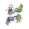
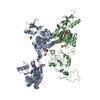
- PDB-3gei: Crystal structure of MnmE from Chlorobium tepidum in complex with GCP -
+ Open data

- Basic information
| Entry | Database: PDB / ID: 3gei | ||||||
|---|---|---|---|---|---|---|---|
| Title | Crystal structure of MnmE from Chlorobium tepidum in complex with GCP | ||||||
 Components Components | tRNA modification GTPase mnmE | ||||||
 Keywords Keywords | HYDROLASE / G protein / G domain / GTPase / GidA / tRNA modification / U34 / GTP-binding / THF-binding / Cytoplasm / Magnesium / Metal-binding / Nucleotide-binding / Potassium / tRNA processing' | ||||||
| Function / homology |  Function and homology information Function and homology informationHydrolases; Acting on acid anhydrides / tRNA wobble uridine modification / tRNA methylation / GTPase activity / GTP binding / metal ion binding / identical protein binding / cytosol Similarity search - Function | ||||||
| Biological species |  Chlorobium tepidum (bacteria) Chlorobium tepidum (bacteria) | ||||||
| Method |  X-RAY DIFFRACTION / SYNCHROTRON / MOLECULAR REPLACEMENT / Resolution: 3.4 Å X-RAY DIFFRACTION / SYNCHROTRON / MOLECULAR REPLACEMENT / Resolution: 3.4 Å | ||||||
 Authors Authors | Meyer, S. / Wittinghofer, A. | ||||||
 Citation Citation | Journal: Plos Biol. / Year: 2009 Title: Kissing G domains of MnmE monitored by X-ray crystallography and pulse electron paramagnetic resonance spectroscopy Authors: Meyer, S. / Bohme, S. / Kruger, A. / Steinhoff, H.-J. / Klare, J.P. / Wittinghofer, A. | ||||||
| History |
|
- Structure visualization
| Structure viewer | Molecule: MolmilJmol/JSmol |
|---|
- Downloads & links
-Download
| PDBx/mmCIF format | 3gei.cif.gz | 228.1 KB | Display | PDBx/mmCIF format |
|---|---|---|---|---|
| PDB format | pdb3gei.ent.gz | 179.4 KB | Display | PDB format |
| PDBx/mmJSON format | 3gei.json.gz | Tree view | PDBx/mmJSON format | |
| Others |  Other downloads Other downloads |
-Validation report
| Arichive directory | https://data.pdbj.org/pub/pdb/validation_reports/ge/3geiftp://data.pdbj.org/pub/pdb/validation_reports/ge/3gei | HTTPS FTP |
|---|
-Related structure data
| Related structure data |  3geeC  3gehC  1xzpS S: Starting model for refinement C: citing same article ( |
|---|---|
| Similar structure data |


-Links
 PDBj
PDBj
- Assembly
| Deposited unit | 
| |||||||||||||||||||||||||||||||||||||||||||||||||||||||||||||||||||||||||||||||||||||||||||||||||||||||||||||||||||||||||||||||||||||||||||||||||||||
|---|---|---|---|---|---|---|---|---|---|---|---|---|---|---|---|---|---|---|---|---|---|---|---|---|---|---|---|---|---|---|---|---|---|---|---|---|---|---|---|---|---|---|---|---|---|---|---|---|---|---|---|---|---|---|---|---|---|---|---|---|---|---|---|---|---|---|---|---|---|---|---|---|---|---|---|---|---|---|---|---|---|---|---|---|---|---|---|---|---|---|---|---|---|---|---|---|---|---|---|---|---|---|---|---|---|---|---|---|---|---|---|---|---|---|---|---|---|---|---|---|---|---|---|---|---|---|---|---|---|---|---|---|---|---|---|---|---|---|---|---|---|---|---|---|---|---|---|---|---|---|
| 1 | 
| |||||||||||||||||||||||||||||||||||||||||||||||||||||||||||||||||||||||||||||||||||||||||||||||||||||||||||||||||||||||||||||||||||||||||||||||||||||
| 2 | 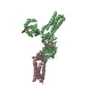
| |||||||||||||||||||||||||||||||||||||||||||||||||||||||||||||||||||||||||||||||||||||||||||||||||||||||||||||||||||||||||||||||||||||||||||||||||||||
| Unit cell |
| |||||||||||||||||||||||||||||||||||||||||||||||||||||||||||||||||||||||||||||||||||||||||||||||||||||||||||||||||||||||||||||||||||||||||||||||||||||
| Noncrystallographic symmetry (NCS) | NCS domain:
NCS domain segments: Refine code: 1
NCS ensembles :
|
-Components
| #1: Protein | Mass: 52165.285 Da / Num. of mol.: 3 Source method: isolated from a genetically manipulated source Source: (gene. exp.) Chlorobium tepidum (bacteria) / Gene: mnmE, CT2084, thdF, trmE / Plasmid: pET14b-CtMnmE / Production host: References: UniProt: Q8KAS1, Hydrolases; Acting on acid anhydrides #2: Chemical |   Mass: 521.208 Da / Num. of mol.: 2 / Source method: obtained synthetically / Formula: C11H18N5O13P3 / Comment: GMP-PCP, energy-carrying molecule analogue*YM Mass: 521.208 Da / Num. of mol.: 2 / Source method: obtained synthetically / Formula: C11H18N5O13P3 / Comment: GMP-PCP, energy-carrying molecule analogue*YM#3: Chemical | ChemComp-MG / |   Mass: 24.305 Da / Num. of mol.: 1 / Source method: obtained synthetically / Formula: Mg Mass: 24.305 Da / Num. of mol.: 1 / Source method: obtained synthetically / Formula: Mg |
|---|
-Experimental details
-Experiment
| Experiment | Method: X-RAY DIFFRACTION / Number of used crystals: 1 |
|---|
- Sample preparation
| Crystal | Density Matthews: 3.93 Å3/Da / Density % sol: 68.73 % |
|---|---|
| Crystal grow | Temperature: 293 K / Method: vapor diffusion, hanging drop / pH: 6 Details: 100mM MES, 46mM NaOH, 12% PEG 4000, 40mM NaCl, 5mM GCP, pH 6.0, VAPOR DIFFUSION, HANGING DROP, temperature 293K |
-Data collection
| Diffraction | Mean temperature: 100 K |
|---|---|
| Diffraction source | Source: SYNCHROTRON / Site: SLS  / Beamline: X10SA / Wavelength: 0.9796 Å / Beamline: X10SA / Wavelength: 0.9796 Å |
| Detector | Type: MAR CCD 165 mm / Detector: CCD / Date: Oct 20, 2006 / Details: Mirrors |
| Radiation | Protocol: SINGLE WAVELENGTH / Monochromatic (M) / Laue (L): M / Scattering type: x-ray |
| Radiation wavelength | Wavelength: 0.9796 Å / Relative weight: 1 |
| Reflection | Resolution: 3.4→20 Å / Num. all: 34300 / Num. obs: 34042 / % possible obs: 99.2 % / Redundancy: 7.45 % / Biso Wilson estimate: 92.53 Å2 / Rmerge(I) obs: 0.129 / Rsym value: 0.131 / Net I/σ(I): 12.13 |
| Reflection shell | Resolution: 3.4→3.425 Å / Redundancy: 7.61 % / Rmerge(I) obs: 0.654 / Mean I/σ(I) obs: 2.02 / Num. unique all: 719 / % possible all: 100 |
- Processing
| Software |
| ||||||||||||||||||||||||||||||||||||||||||||||||||||||||||||||||||||||||||||||||||||||||||||||||||||||||||||||||||||||||||||||||||||||||||||||||||||||||||||||||||||||||||
|---|---|---|---|---|---|---|---|---|---|---|---|---|---|---|---|---|---|---|---|---|---|---|---|---|---|---|---|---|---|---|---|---|---|---|---|---|---|---|---|---|---|---|---|---|---|---|---|---|---|---|---|---|---|---|---|---|---|---|---|---|---|---|---|---|---|---|---|---|---|---|---|---|---|---|---|---|---|---|---|---|---|---|---|---|---|---|---|---|---|---|---|---|---|---|---|---|---|---|---|---|---|---|---|---|---|---|---|---|---|---|---|---|---|---|---|---|---|---|---|---|---|---|---|---|---|---|---|---|---|---|---|---|---|---|---|---|---|---|---|---|---|---|---|---|---|---|---|---|---|---|---|---|---|---|---|---|---|---|---|---|---|---|---|---|---|---|---|---|---|---|---|
| Refinement | Method to determine structure: MOLECULAR REPLACEMENT Starting model: PDB ENTRY 1XZP Resolution: 3.4→19.94 Å / Cor.coef. Fo:Fc: 0.926 / Cor.coef. Fo:Fc free: 0.912 / SU B: 61.478 / SU ML: 0.438 / Cross valid method: THROUGHOUT / ESU R Free: 0.486 / Stereochemistry target values: MAXIMUM LIKELIHOOD / Details: HYDROGENS HAVE BEEN ADDED IN THE RIDING POSITIONS
| ||||||||||||||||||||||||||||||||||||||||||||||||||||||||||||||||||||||||||||||||||||||||||||||||||||||||||||||||||||||||||||||||||||||||||||||||||||||||||||||||||||||||||
| Solvent computation | Ion probe radii: 0.8 Å / Shrinkage radii: 0.8 Å / VDW probe radii: 1.2 Å / Solvent model: MASK | ||||||||||||||||||||||||||||||||||||||||||||||||||||||||||||||||||||||||||||||||||||||||||||||||||||||||||||||||||||||||||||||||||||||||||||||||||||||||||||||||||||||||||
| Displacement parameters | Biso mean: 123.559 Å2
| ||||||||||||||||||||||||||||||||||||||||||||||||||||||||||||||||||||||||||||||||||||||||||||||||||||||||||||||||||||||||||||||||||||||||||||||||||||||||||||||||||||||||||
| Refinement step | Cycle: LAST / Resolution: 3.4→19.94 Å
| ||||||||||||||||||||||||||||||||||||||||||||||||||||||||||||||||||||||||||||||||||||||||||||||||||||||||||||||||||||||||||||||||||||||||||||||||||||||||||||||||||||||||||
| Refine LS restraints |
| ||||||||||||||||||||||||||||||||||||||||||||||||||||||||||||||||||||||||||||||||||||||||||||||||||||||||||||||||||||||||||||||||||||||||||||||||||||||||||||||||||||||||||
| Refine LS restraints NCS | Refine-ID: X-RAY DIFFRACTION
| ||||||||||||||||||||||||||||||||||||||||||||||||||||||||||||||||||||||||||||||||||||||||||||||||||||||||||||||||||||||||||||||||||||||||||||||||||||||||||||||||||||||||||
| LS refinement shell | Resolution: 3.4→3.488 Å / Total num. of bins used: 20
|
