 Movie
Movie Controller

[English] 日本語
 Yorodumi
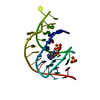
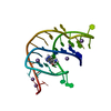
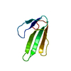
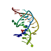
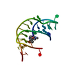
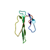
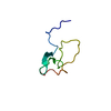
- PDB-3gca: The structural basis for recognition of the preQ0 metabolite by a... -
+ Open data

- Basic information
| Entry | Database: PDB / ID: 3gca | ||||||
|---|---|---|---|---|---|---|---|
| Title | The structural basis for recognition of the preQ0 metabolite by an unusually small riboswitch aptamer domain | ||||||
 Components Components | PreQ1 riboswitch | ||||||
 Keywords Keywords | RNA / PreQ1 / PreQ0 / riboswitch / ribosomal binding site / amptamer / metabolite | ||||||
| Function / homology | Chem-PQ0 / RNA / RNA (> 10) Function and homology information Function and homology information | ||||||
| Method |  X-RAY DIFFRACTION / SYNCHROTRON / MAD / Resolution: 2.75 Å X-RAY DIFFRACTION / SYNCHROTRON / MAD / Resolution: 2.75 Å | ||||||
 Authors Authors | Spitale, R.C. / Wedekind, J.E. | ||||||
 Citation Citation | Journal: J.Biol.Chem. / Year: 2009 Title: The Structural Basis for Recognition of the PreQ0 Metabolite by an Unusually Small Riboswitch Aptamer Domain. Authors: Spitale, R.C. / Torelli, A.T. / Krucinska, J. / Bandarian, V. / Wedekind, J.E. | ||||||
| History |
|
- Structure visualization
| Structure viewer | Molecule: MolmilJmol/JSmol |
|---|
- Downloads & links
-Download
| PDBx/mmCIF format | 3gca.cif.gz | 27.7 KB | Display | PDBx/mmCIF format |
|---|---|---|---|---|
| PDB format | pdb3gca.ent.gz | 19.3 KB | Display | PDB format |
| PDBx/mmJSON format | 3gca.json.gz | Tree view | PDBx/mmJSON format | |
| Others |  Other downloads Other downloads |
-Validation report
| Arichive directory | https://data.pdbj.org/pub/pdb/validation_reports/gc/3gcaftp://data.pdbj.org/pub/pdb/validation_reports/gc/3gca | HTTPS FTP |
|---|
-Related structure data
| Similar structure data |
|---|


-Links
 PDBj
PDBj


- Assembly
| Deposited unit | 
| ||||||||
|---|---|---|---|---|---|---|---|---|---|
| 1 |
| ||||||||
| Unit cell |
|
-Components
| #1: RNA chain | Mass: 10599.417 Da / Num. of mol.: 1 / Source method: obtained synthetically Details: The RNA strand was chemically synthesized based on the sequence of Class I, Type I PreQ1 riboswitch aptamer domain from T. tengcongensis |
|---|---|
| #2: Chemical | ChemComp-PQ0 / 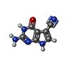  Mass: 175.147 Da / Num. of mol.: 1 / Source method: obtained synthetically / Formula: C7H5N5O Mass: 175.147 Da / Num. of mol.: 1 / Source method: obtained synthetically / Formula: C7H5N5O |
| #3: Chemical |   Mass: 96.063 Da / Num. of mol.: 2 / Source method: obtained synthetically / Formula: SO4 Mass: 96.063 Da / Num. of mol.: 2 / Source method: obtained synthetically / Formula: SO4 |
-Experimental details
-Experiment
| Experiment | Method: X-RAY DIFFRACTION / Number of used crystals: 1 |
|---|
- Sample preparation
| Crystal | Density Matthews: 4.93 Å3/Da / Density % sol: 75.05 % | ||||||||||||||||||||||||||||||||||||||||
|---|---|---|---|---|---|---|---|---|---|---|---|---|---|---|---|---|---|---|---|---|---|---|---|---|---|---|---|---|---|---|---|---|---|---|---|---|---|---|---|---|---|
| Crystal grow | Temperature: 293 K / Method: vapor diffusion, hanging drop / pH: 6 Details: 1.8 M Li2SO4, 0.10 M Na-cacodylate pH 6.0, 0.01 M Mg(SO4)2-, 5% (v/v) 1,3-propanediol and 2 mM spermine., VAPOR DIFFUSION, HANGING DROP, temperature 293K | ||||||||||||||||||||||||||||||||||||||||
| Components of the solutions |
|
-Data collection
| Diffraction | Mean temperature: 100 K | ||||||||||||
|---|---|---|---|---|---|---|---|---|---|---|---|---|---|
| Diffraction source | Source: SYNCHROTRON / Site: SSRL  / Beamline: BL7-1 / Wavelength: 1.1395, 1.1407, 0.97622, 1.1395 / Beamline: BL7-1 / Wavelength: 1.1395, 1.1407, 0.97622, 1.1395 | ||||||||||||
| Detector | Type: Quantum 315 CCD detector / Detector: CCD / Date: Jan 29, 2009 Details: Vertical focusing mirror; single crystal (Si111) bent monochromator (horizontal focusing). | ||||||||||||
| Radiation | Protocol: MAD / Monochromatic (M) / Laue (L): M / Scattering type: x-ray | ||||||||||||
| Radiation wavelength |
| ||||||||||||
| Reflection | Resolution: 2.75→50 Å / Num. obs: 5990 / % possible obs: 97.6 % / Observed criterion σ(F): 0 / Observed criterion σ(I): -3 / Redundancy: 16.6 % / Biso Wilson estimate: 75 Å2 / Rsym value: 0.056 / Net I/σ(I): 36.47 | ||||||||||||
| Reflection shell | Resolution: 2.75→2.85 Å / Redundancy: 15.8 % / Mean I/σ(I) obs: 6.5 / Num. unique all: 579 / Rsym value: 0.36 / % possible all: 98.5 |
- Processing
| Software |
| |||||||||||||||||||||
|---|---|---|---|---|---|---|---|---|---|---|---|---|---|---|---|---|---|---|---|---|---|---|
| Refinement | Method to determine structure: MAD / Resolution: 2.75→28.33 Å / Rfactor Rfree error: 0.013 / Data cutoff high absF: 303382.6 / Data cutoff low absF: 0 / Isotropic thermal model: RESTRAINED / Cross valid method: THROUGHOUT / σ(F): 0 / Stereochemistry target values: CNS 1.2 / Details: BULK SOLVENT MODEL USED
| |||||||||||||||||||||
| Solvent computation | Solvent model: FLAT MODEL / Bsol: 51.0924 Å2 / ksol: 0.2 e/Å3 | |||||||||||||||||||||
| Displacement parameters | Biso mean: 76 Å2
| |||||||||||||||||||||
| Refine analyze |
| |||||||||||||||||||||
| Refinement step | Cycle: LAST / Resolution: 2.75→28.33 Å
| |||||||||||||||||||||
| Refine LS restraints |
| |||||||||||||||||||||
| LS refinement shell | Resolution: 2.75→2.92 Å / Rfactor Rfree error: 0.06 / Total num. of bins used: 6
| |||||||||||||||||||||
| Xplor file |
|
