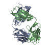
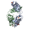
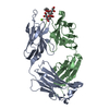
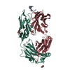
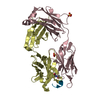
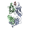
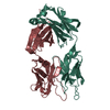
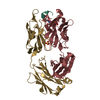
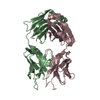
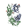
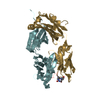
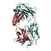
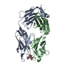
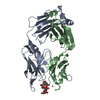
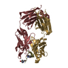
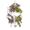
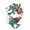
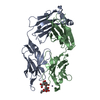
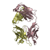
Entry Database : PDB / ID : 3g5yTitle Antibodies Specifically Targeting a Locally Misfolded Region of Tumor Associated EGFR 175 heavy chain 175 light chain EGFR peptide Keywords / Function / homology Function Domain/homology Component
/ / / / / / / / / / / / / / / / / / / / / / / / / / / / / / / / / / / / / / / / / / / / / / / / / / / / / / / / / / / / / / / / / / / / / / / / / / / / / / / / / / / / / / / / / / / / / / / / / / / / / / / / / / / / / / / / / / / / / / / / / / / / / / / / / / / / / / / / / / / / / / / / / Biological species Mus musculus (house mouse)Method / / Resolution : 1.59 Å Authors Garrett, T.P.J. / Burgess, A.W. Journal : Proc.Natl.Acad.Sci.USA / Year : 2009Title : Antibodies specifically targeting a locally misfolded region of tumor associated EGFRAuthors: Garrett, T.P.J. / Burgess, A.W. / Gan, H.K. / Luwor, R.B. / Cartwright, G. / Walker, F. / Orchard, S.G. / Clayton, A.H.A. / Nice, E.C. / Rothacker, J. / Catimel, B. / Cavenee, W.K. / Old, L. ... Authors : Garrett, T.P.J. / Burgess, A.W. / Gan, H.K. / Luwor, R.B. / Cartwright, G. / Walker, F. / Orchard, S.G. / Clayton, A.H.A. / Nice, E.C. / Rothacker, J. / Catimel, B. / Cavenee, W.K. / Old, L.J. / Stockert, E. / Ritter, G. / Adams, T.E. / Hoyne, P.A. / Wittrup, D. / Chao, G. / Cochran, J.R. / Luo, C. / Lou, M. / Huyton, T. / Xu, Y. / Fairlie, W.D. / Yao, S. / Scott, A.M. / Johns, T.G. History Deposition Feb 5, 2009 Deposition site / Processing site Revision 1.0 Feb 9, 2010 Provider / Type Revision 1.1 Jul 13, 2011 Group / Version format complianceRevision 1.2 Oct 12, 2011 Group Revision 1.3 Oct 16, 2024 Group / Database references / Structure summaryCategory chem_comp_atom / chem_comp_bond ... chem_comp_atom / chem_comp_bond / database_2 / pdbx_entry_details / pdbx_modification_feature Item / _database_2.pdbx_database_accession / _pdbx_entry_details.has_protein_modification
Show all Show less
 Movie
Movie Controller

 Yorodumi
 Open data
 Basic information
 Components
 Keywords
 Function and homology information
 X-RAY DIFFRACTION /
  Authors
 Citation
 Structure visualization
 Downloads & links
 Other downloads
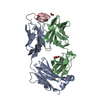


 PDBj
PDBj


 Assembly
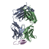
 Mass: 18.015 Da / Num. of mol.: 247 / Source method: isolated from a natural source / Formula: H2O
 Sample preparation
 Processing