- PDB-3fu1: Crystal structure of the major pseudopilin from the type 2 secret... -
+
Open data
ID or keywords:
Loading...
-
Basic information
Entry
Database: PDB / ID: 3fu1
Title
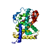
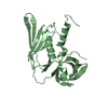
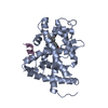
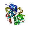
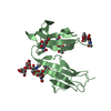
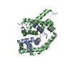
Crystal structure of the major pseudopilin from the type 2 secretion system of Vibrio cholerae
Components
General secretion pathway protein G
Keywords
PROTEIN TRANSPORT / GENERAL SECRETORY PATHWAY / MAJOR PILIN / COMPLEX / Methylation / Transport
Function / homology
Function and homology information
protein secretion by the type II secretion system / type II protein secretion system complex / plasma membrane Similarity search - Function
Type II secretion system protein GspG / Type II secretion system protein GspG, C-terminal / Type II secretion system (T2SS), protein G / Glycoprotein, Type 4 Pilin / Bacterial general secretion pathway protein G-type pilin / Glycoprotein, Type 4 Pilin / : / Prokaryotic N-terminal methylation site. / Prokaryotic N-terminal methylation motif / Prokaryotic N-terminal methylation site ...Type II secretion system protein GspG / Type II secretion system protein GspG, C-terminal / Type II secretion system (T2SS), protein G / Glycoprotein, Type 4 Pilin / Bacterial general secretion pathway protein G-type pilin / Glycoprotein, Type 4 Pilin / : / Prokaryotic N-terminal methylation site. / Prokaryotic N-terminal methylation motif / Prokaryotic N-terminal methylation site / Pilin-like / 2-Layer Sandwich / Alpha Beta Similarity search - Domain/homology
THIS CRYSTAL FORM CONTAINS A DIMER, THAT IS NOT RELATED TO THE BIOLOGICAL FORM. BIOLOGICAL UNIT IS A FIBER, IT IS UNKNOWN AT THE MOMENT HOW INDIVIDUAL SUBUNITS ARE ASSEMBLED IN VIVO.
-
Components
#1: Protein
GeneralsecretionpathwayproteinG / Cholera toxin secretion protein epsG
Mass: 12579.704 Da / Num. of mol.: 2 / Fragment: UNP RESIDUES 35-146 Source method: isolated from a genetically manipulated source Details: RESIDUES 26-137 / Source: (gene. exp.) Vibrio cholerae (bacteria) / Strain: 569B / Gene: epsG, VC_2730 / Plasmid: pCDF-NT / Production host: Escherichia coli (E. coli) / Strain (production host): BL21(DE3) / References: UniProt: P45773
Mass: 18.015 Da / Num. of mol.: 224 / Source method: isolated from a natural source / Formula: H2O
-
Experimental details
-
Experiment
Experiment
Method: X-RAY DIFFRACTION / Number of used crystals: 1
-
Sample preparation
Crystal
Density Matthews: 2.12 Å3/Da / Density % sol: 41.95 %
Crystal grow
Temperature: 298 K / Method: vapor diffusion, sitting drop / pH: 5 Details: 22.5% PEG 3350, 0.1M NA ACETATE, 0.04M ZN ACETATE, pH 5.0, vapor diffusion, sitting drop, temperature 298K
-
Data collection
Diffraction
ID
Mean temperature (K)
Crystal-ID
1
100
1
2
100
1
1,2
1
Diffraction source
Source
Site
Beamline
ID
Wavelength (Å)
SYNCHROTRON
SSRL
BL9-2
1
0.97946
SYNCHROTRON
SSRL
BL9-2
2
1.28344
Detector
Type
ID
Detector
Date
MARMOSAIC 325 mm CCD
1
CCD
Nov 15, 2007
MARMOSAIC 325 mm CCD
2
CCD
Nov 15, 2007
Radiation
ID
Monochromator
Protocol
Monochromatic (M) / Laue (L)
Scattering type
Wavelength-ID
1
DOUBLECRYSTALSI(111)
SINGLEWAVELENGTH
M
x-ray
1
2
DOUBLECRYSTALSI(111)
SINGLEWAVELENGTH
M
x-ray
2
Radiation wavelength
ID
Wavelength (Å)
Relative weight
1
0.97946
1
2
1.28344
1
Reflection
Redundancy: 6.7 % / Av σ(I) over netI: 24.02 / Number: 113449 / Rmerge(I) obs: 0.087 / Χ2: 1.43 / D res high: 1.9 Å / D res low: 29 Å / Num. obs: 17045 / % possible obs: 97.1
Diffraction reflection shell
Highest resolution (Å)
Lowest resolution (Å)
% possible obs (%)
ID
Rmerge(I) obs
Chi squared
Redundancy
4.09
29
99.9
1
0.04
1.896
6.8
3.25
4.09
100
1
0.048
1.673
7.2
2.84
3.25
100
1
0.087
1.593
7.3
2.58
2.84
100
1
0.132
1.331
7.3
2.39
2.58
100
1
0.174
1.271
7.3
2.25
2.39
100
1
0.229
1.281
7.3
2.14
2.25
99.7
1
0.291
1.289
7
2.05
2.14
98.1
1
0.371
1.245
6.3
1.97
2.05
93.5
1
0.478
1.232
5.3
1.9
1.97
78.8
1
0.512
1.226
4.1
Reflection
Resolution: 1.9→29 Å / Num. obs: 17045 / % possible obs: 97.1 % / Redundancy: 6.7 % / Biso Wilson estimate: 24 Å2 / Rmerge(I) obs: 0.087 / Χ2: 1.428 / Net I/σ(I): 24.016
Reflection shell
Resolution: 1.9→1.97 Å / Redundancy: 4.1 % / Rmerge(I) obs: 0.512 / Mean I/σ(I) obs: 2.1 / Num. unique all: 1373 / Χ2: 1.226 / % possible all: 78.8
-
Phasing
Phasing
Method: molecular replacement
Phasing MR
Model details: Phaser MODE: MR_AUTO
Highest resolution
Lowest resolution
Rotation
2.5 Å
29.04 Å
Translation
2.5 Å
29.04 Å
-
Processing
Software
Name
Version
Classification
NB
DENZO
datareduction
SCALEPACK
datascaling
PHASER
phasing
REFMAC
refinement
PDB_EXTRACT
3.006
dataextraction
Blu-Ice
datacollection
HKL-2000
datareduction
HKL-2000
datascaling
Refinement
Method to determine structure: MOLECULAR REPLACEMENT / Resolution: 1.9→27.28 Å / Cor.coef. Fo:Fc: 0.958 / Cor.coef. Fo:Fc free: 0.932 / WRfactor Rfree: 0.212 / WRfactor Rwork: 0.166 / Occupancy max: 1 / Occupancy min: 0 / FOM work R set: 0.875 / SU B: 6.78 / SU ML: 0.101 / SU R Cruickshank DPI: 0.172 / SU Rfree: 0.153 / TLS residual ADP flag: LIKELY RESIDUAL / Cross valid method: THROUGHOUT / σ(F): 0 / ESU R: 0.172 / ESU R Free: 0.153 / Stereochemistry target values: MAXIMUM LIKELIHOOD Details: HYDROGENS HAVE BEEN ADDED IN THE RIDING POSITIONS. U VALUES: RESIDUAL ONLY
Rfactor
Num. reflection
% reflection
Selection details
Rfree
0.223
830
4.9 %
RANDOM
Rwork
0.178
-
-
-
obs
0.18
16995
96.88 %
-
Solvent computation
Ion probe radii: 0.8 Å / Shrinkage radii: 0.8 Å / VDW probe radii: 1.2 Å / Solvent model: BABINET MODEL WITH MASK
In the structure databanks used in Yorodumi, some data are registered as the other names, "COVID-19 virus" and "2019-nCoV". Here are the details of the virus and the list of structure data.
Jan 31, 2019. EMDB accession codes are about to change! (news from PDBe EMDB page)
EMDB accession codes are about to change! (news from PDBe EMDB page)
The allocation of 4 digits for EMDB accession codes will soon come to an end. Whilst these codes will remain in use, new EMDB accession codes will include an additional digit and will expand incrementally as the available range of codes is exhausted. The current 4-digit format prefixed with “EMD-” (i.e. EMD-XXXX) will advance to a 5-digit format (i.e. EMD-XXXXX), and so on. It is currently estimated that the 4-digit codes will be depleted around Spring 2019, at which point the 5-digit format will come into force.
The EM Navigator/Yorodumi systems omit the EMD- prefix.
Related info.:Q: What is EMD? / ID/Accession-code notation in Yorodumi/EM Navigator
Yorodumi is a browser for structure data from EMDB, PDB, SASBDB, etc.
This page is also the successor to EM Navigator detail page, and also detail information page/front-end page for Omokage search.
The word "yorodu" (or yorozu) is an old Japanese word meaning "ten thousand". "mi" (miru) is to see.
Related info.:EMDB / PDB / SASBDB / Comparison of 3 databanks / Yorodumi Search / Aug 31, 2016. New EM Navigator & Yorodumi / Yorodumi Papers / Jmol/JSmol / Function and homology information / Changes in new EM Navigator and Yorodumi
 Movie
Movie Controller

 Yorodumi
 Open data
 Basic information
 Components
 Keywords
 Function and homology information
 Vibrio cholerae (bacteria)
 X-RAY DIFFRACTION /
  Authors
 Citation
 Structure visualization
 Downloads & links
 Other downloads
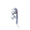


 PDBj
PDBj
 Assembly
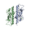

 Mass: 65.409 Da / Num. of mol.: 3 / Source method: obtained synthetically / Formula: Zn
 Mass: 40.078 Da / Num. of mol.: 2 / Source method: obtained synthetically / Formula: Ca
 Mass: 18.015 Da / Num. of mol.: 224 / Source method: isolated from a natural source / Formula: H2O
 Sample preparation
 Processing