- PDB-3g20: Crystal structure of the major pseudopilin from the type 2 secret... -
+
Open data
ID or keywords:
Loading...
-
Basic information
Entry
Database: PDB / ID: 3g20
Title
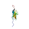
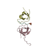
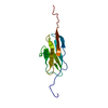
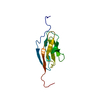
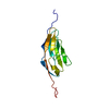
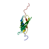
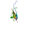
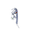
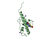
Crystal structure of the major pseudopilin from the type 2 secretion system of enterohaemorrhagic Escherichia coli
Components
Type II secretion protein
Keywords
PROTEIN TRANSPORT / GENERAL SECRETORY PATHWAY / MAJOR PILIN / COMPLEX
Function / homology
Function and homology information
protein secretion by the type II secretion system / type II protein secretion system complex / metal ion binding / identical protein binding / plasma membrane Similarity search - Function
Type II secretion system protein GspG / Type II secretion system protein GspG, C-terminal / Type II secretion system (T2SS), protein G / Glycoprotein, Type 4 Pilin / Bacterial general secretion pathway protein G-type pilin / Glycoprotein, Type 4 Pilin / Prokaryotic N-terminal methylation site. / Prokaryotic N-terminal methylation motif / Prokaryotic N-terminal methylation site / Pilin-like ...Type II secretion system protein GspG / Type II secretion system protein GspG, C-terminal / Type II secretion system (T2SS), protein G / Glycoprotein, Type 4 Pilin / Bacterial general secretion pathway protein G-type pilin / Glycoprotein, Type 4 Pilin / Prokaryotic N-terminal methylation site. / Prokaryotic N-terminal methylation motif / Prokaryotic N-terminal methylation site / Pilin-like / 2-Layer Sandwich / Alpha Beta Similarity search - Domain/homology
THIS CRYSTAL FORM CONTAINS TWO MONOMERS THAT ARE NOT RELATED TO THE BIOLOGICAL FORM. THE BIOLOGICAL UNIT IS A FIBER. IT IS UNKNOWN AT THE MOMENT HOW INDIVIDUAL SUBUNITS ARE ASSEMBLED IN VIVO.
-
Components
-
Protein , 1 types, 2 molecules AB
#1: Protein
TypeIIsecretionprotein / Type II secretion pathway related protein
Mass: 13733.197 Da / Num. of mol.: 2 / Fragment: UNP residues 24-144 Source method: isolated from a genetically manipulated source Source: (gene. exp.) Escherichia coli O157:H7 (bacteria) / Strain: 86-24 / Gene: ECO57PM05, etpG, GSPG, L7036 / Plasmid: pCDF-NT / Production host: Escherichia coli (E. coli) / Strain (production host): BL21(DE3) / References: UniProt: Q7BSV8
In the structure databanks used in Yorodumi, some data are registered as the other names, "COVID-19 virus" and "2019-nCoV". Here are the details of the virus and the list of structure data.
Jan 31, 2019. EMDB accession codes are about to change! (news from PDBe EMDB page)
EMDB accession codes are about to change! (news from PDBe EMDB page)
The allocation of 4 digits for EMDB accession codes will soon come to an end. Whilst these codes will remain in use, new EMDB accession codes will include an additional digit and will expand incrementally as the available range of codes is exhausted. The current 4-digit format prefixed with “EMD-” (i.e. EMD-XXXX) will advance to a 5-digit format (i.e. EMD-XXXXX), and so on. It is currently estimated that the 4-digit codes will be depleted around Spring 2019, at which point the 5-digit format will come into force.
The EM Navigator/Yorodumi systems omit the EMD- prefix.
Related info.:Q: What is EMD? / ID/Accession-code notation in Yorodumi/EM Navigator
Yorodumi is a browser for structure data from EMDB, PDB, SASBDB, etc.
This page is also the successor to EM Navigator detail page, and also detail information page/front-end page for Omokage search.
The word "yorodu" (or yorozu) is an old Japanese word meaning "ten thousand". "mi" (miru) is to see.
Related info.:EMDB / PDB / SASBDB / Comparison of 3 databanks / Yorodumi Search / Aug 31, 2016. New EM Navigator & Yorodumi / Yorodumi Papers / Jmol/JSmol / Function and homology information / Changes in new EM Navigator and Yorodumi
 Movie
Movie Controller

 Yorodumi
 Open data
 Basic information
 Components
 Keywords
 Function and homology information
 X-RAY DIFFRACTION /
  Authors
 Citation
 Structure visualization
 Downloads & links
 Other downloads
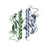


 PDBj
PDBj
 Assembly
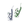


 Mass: 207.290 Da / Num. of mol.: 2 / Source method: obtained synthetically / Formula: C8H17NO3S / Comment: pH buffer*YM
 Mass: 40.078 Da / Num. of mol.: 2 / Source method: obtained synthetically / Formula: Ca
 Mass: 22.990 Da / Num. of mol.: 3 / Source method: obtained synthetically / Formula: Na
 Mass: 92.094 Da / Num. of mol.: 1 / Source method: obtained synthetically / Formula: C3H8O3
 Sample preparation
 / Beamline: BL9-2 / Wavelength: 0.97945 Å
 Processing