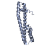
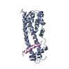
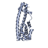
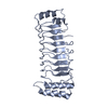
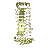
- PDB-3frr: Structure of human IST1(NTD) - (residues 1-189)(P21) -
+
Open data
ID or keywords:
Loading...
-
Basic information
Entry
Database: PDB / ID: 3frr
Title
Structure of human IST1(NTD) - (residues 1-189)(P21)
Components
Uncharacterized protein KIAA0174
Keywords
PROTEIN BINDING / ESCRT / ESCRT-III / CHMP / IST1 / Alternative splicing / Phosphoprotein
Function / homology
Function and homology information
MIT domain binding / ESCRT III complex disassembly / cytoskeleton-dependent cytokinesis / collateral sprouting / positive regulation of collateral sprouting / Sealing of the nuclear envelope (NE) by ESCRT-III / midbody abscission / multivesicular body assembly / Flemming body / endoplasmic reticulum-Golgi intermediate compartment ...MIT domain binding / ESCRT III complex disassembly / cytoskeleton-dependent cytokinesis / collateral sprouting / positive regulation of collateral sprouting / Sealing of the nuclear envelope (NE) by ESCRT-III / midbody abscission / multivesicular body assembly / Flemming body / endoplasmic reticulum-Golgi intermediate compartment / positive regulation of proteolysis / establishment of protein localization / azurophil granule lumen / intracellular protein localization / nuclear envelope / protein transport / midbody / cadherin binding / protein domain specific binding / cell division / intracellular membrane-bounded organelle / centrosome / Neutrophil degranulation / protein-containing complex binding / chromatin / extracellular exosome / extracellular region / identical protein binding / cytosol Similarity search - Function
Vacuolar protein sorting-associated protein Ist1 / Vacuolar protein sorting-associated protein Ist1 / Vacuolar protein sorting-associated protein IST1-like / Regulator of Vps4 activity in the MVB pathway / Ferritin / Up-down Bundle / Mainly Alpha Similarity search - Domain/homology
In the structure databanks used in Yorodumi, some data are registered as the other names, "COVID-19 virus" and "2019-nCoV". Here are the details of the virus and the list of structure data.
Jan 31, 2019. EMDB accession codes are about to change! (news from PDBe EMDB page)
EMDB accession codes are about to change! (news from PDBe EMDB page)
The allocation of 4 digits for EMDB accession codes will soon come to an end. Whilst these codes will remain in use, new EMDB accession codes will include an additional digit and will expand incrementally as the available range of codes is exhausted. The current 4-digit format prefixed with “EMD-” (i.e. EMD-XXXX) will advance to a 5-digit format (i.e. EMD-XXXXX), and so on. It is currently estimated that the 4-digit codes will be depleted around Spring 2019, at which point the 5-digit format will come into force.
The EM Navigator/Yorodumi systems omit the EMD- prefix.
Related info.:Q: What is EMD? / ID/Accession-code notation in Yorodumi/EM Navigator
Yorodumi is a browser for structure data from EMDB, PDB, SASBDB, etc.
This page is also the successor to EM Navigator detail page, and also detail information page/front-end page for Omokage search.
The word "yorodu" (or yorozu) is an old Japanese word meaning "ten thousand". "mi" (miru) is to see.
Related info.:EMDB / PDB / SASBDB / Comparison of 3 databanks / Yorodumi Search / Aug 31, 2016. New EM Navigator & Yorodumi / Yorodumi Papers / Jmol/JSmol / Function and homology information / Changes in new EM Navigator and Yorodumi
 Movie
Movie Controller


 Open data
 Basic information
 Components
 Keywords
 Function and homology information
 Homo sapiens (human)
 X-RAY DIFFRACTION /
  Authors
 Citation
 Structure visualization
 Downloads & links
 Other downloads


 PDBj
PDBj Assembly
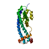

 Mass: 18.015 Da / Num. of mol.: 163 / Source method: isolated from a natural source / Formula: H2O
 Sample preparation
 Processing