 Movie
Movie Controller


+ Open data

- Basic information
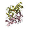
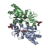
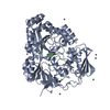
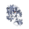
| Entry | Database: PDB / ID: 3fj7 | ||||||
|---|---|---|---|---|---|---|---|
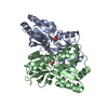
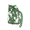
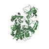
| Title | Crystal structure of L-phospholactate Bound PEB3 | ||||||
 Components Components | Major antigenic peptide PEB3 | ||||||
 Keywords Keywords | PROTEIN BINDING / PEB3 / PEP | ||||||
| Function / homology |  Function and homology information Function and homology informationsulfate transmembrane transport / molecular carrier activity / periplasmic space Similarity search - Function | ||||||
| Biological species |   Campylobacter jejuni (Campylobacter) Campylobacter jejuni (Campylobacter) | ||||||
| Method |  X-RAY DIFFRACTION / SYNCHROTRON / MOLECULAR REPLACEMENT / Resolution: 1.7 Å X-RAY DIFFRACTION / SYNCHROTRON / MOLECULAR REPLACEMENT / Resolution: 1.7 Å | ||||||
 Authors Authors | Min, T. / Matte, A. / Cygler, M. | ||||||
 Citation Citation | Journal: Biochemistry / Year: 2009 Title: Specificity of Campylobacter jejuni adhesin PEB3 for phosphates and structural differences among its ligand complexes. Authors: Min, T. / Vedadi, M. / Watson, D.C. / Wasney, G.A. / Munger, C. / Cygler, M. / Matte, A. / Young, N.M. | ||||||
| History |
|
- Structure visualization
| Structure viewer | Molecule: MolmilJmol/JSmol |
|---|
- Downloads & links
-Download
| PDBx/mmCIF format | 3fj7.cif.gz | 110.2 KB | Display | PDBx/mmCIF format |
|---|---|---|---|---|
| PDB format | pdb3fj7.ent.gz | 83.5 KB | Display | PDB format |
| PDBx/mmJSON format | 3fj7.json.gz | Tree view | PDBx/mmJSON format | |
| Others |  Other downloads Other downloads |
-Validation report
| Arichive directory | https://data.pdbj.org/pub/pdb/validation_reports/fj/3fj7ftp://data.pdbj.org/pub/pdb/validation_reports/fj/3fj7 | HTTPS FTP |
|---|
-Related structure data
| Related structure data |  3firC  3fjgC  3fjmC 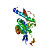 2hwxS C: citing same article ( S: Starting model for refinement |
|---|---|
| Similar structure data |

-Links
 PDBj
PDBj- Assembly
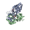
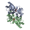
| Deposited unit | 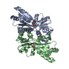
| ||||||||
|---|---|---|---|---|---|---|---|---|---|
| 1 |
| ||||||||
| Unit cell |
|
-Components
| #1: Protein | Mass: 25626.092 Da / Num. of mol.: 2 / Fragment: UNP residues 21-250 Source method: isolated from a genetically manipulated source Source: (gene. exp.) Campylobacter jejuni (Campylobacter) / Strain: 11168 / Gene: cj0289c / Plasmid: pUC18 / Production host: #2: Chemical | 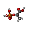  Mass: 170.058 Da / Num. of mol.: 2 / Source method: obtained synthetically / Formula: C3H7O6P Mass: 170.058 Da / Num. of mol.: 2 / Source method: obtained synthetically / Formula: C3H7O6P#3: Water | ChemComp-HOH / |  Mass: 18.015 Da / Num. of mol.: 400 / Source method: isolated from a natural source / Formula: H2O Mass: 18.015 Da / Num. of mol.: 400 / Source method: isolated from a natural source / Formula: H2O |
|---|
-Experimental details
-Experiment
| Experiment | Method: X-RAY DIFFRACTION / Number of used crystals: 1 |
|---|
- Sample preparation
| Crystal | Density Matthews: 2.2 Å3/Da / Density % sol: 44.17 % |
|---|---|
| Crystal grow | Temperature: 294 K / Method: vapor diffusion, hanging drop / pH: 5.5 Details: 18% [w/v] polyethylene glycol 3350, 0.2 M ammonium acetate, pH 5.5, VAPOR DIFFUSION, HANGING DROP, temperature 294K |
-Data collection
| Diffraction | Mean temperature: 100 K |
|---|---|
| Diffraction source | Source: SYNCHROTRON / Site: APS  / Beamline: 31-ID / Wavelength: 0.98 Å / Beamline: 31-ID / Wavelength: 0.98 Å |
| Detector | Type: MARRESEARCH / Detector: CCD / Date: Mar 19, 2008 / Details: Vertical and Horizontal focusing Mirrors |
| Radiation | Monochromator: Diamond / Protocol: SINGLE WAVELENGTH / Monochromatic (M) / Laue (L): M / Scattering type: x-ray |
| Radiation wavelength | Wavelength: 0.98 Å / Relative weight: 1 |
| Reflection | Resolution: 1.7→43.9 Å / Num. obs: 54648 / % possible obs: 100 % / Redundancy: 7.2 % / Biso Wilson estimate: 17 Å2 / Rsym value: 0.083 / Net I/σ(I): 11.6 |
| Reflection shell | Resolution: 1.7→1.79 Å / Redundancy: 7 % / Rmerge(I) obs: 0.266 / Mean I/σ(I) obs: 7.7 / Num. unique all: 7879 / Rsym value: 0.287 / % possible all: 99.9 |
- Processing
| Software |
| |||||||||||||||||||||||||||||||||||||||||||||||||||||||||||||||||
|---|---|---|---|---|---|---|---|---|---|---|---|---|---|---|---|---|---|---|---|---|---|---|---|---|---|---|---|---|---|---|---|---|---|---|---|---|---|---|---|---|---|---|---|---|---|---|---|---|---|---|---|---|---|---|---|---|---|---|---|---|---|---|---|---|---|---|
| Refinement | Method to determine structure: MOLECULAR REPLACEMENT Starting model: PDB ENTRY 2HWX,PEB3 citrate bound form Resolution: 1.7→43.9 Å / Cor.coef. Fo:Fc: 0.935 / Cor.coef. Fo:Fc free: 0.925 / SU B: 2.221 / SU ML: 0.075 / Cross valid method: THROUGHOUT / ESU R: 0.127 / ESU R Free: 0.117 / Stereochemistry target values: MAXIMUM LIKELIHOOD / Details: HYDROGENS HAVE BEEN ADDED IN THE RIDING POSITIONS
| |||||||||||||||||||||||||||||||||||||||||||||||||||||||||||||||||
| Solvent computation | Ion probe radii: 0.8 Å / Shrinkage radii: 0.8 Å / VDW probe radii: 1.2 Å / Solvent model: MASK | |||||||||||||||||||||||||||||||||||||||||||||||||||||||||||||||||
| Displacement parameters | Biso mean: 13.756 Å2
| |||||||||||||||||||||||||||||||||||||||||||||||||||||||||||||||||
| Refinement step | Cycle: LAST / Resolution: 1.7→43.9 Å
| |||||||||||||||||||||||||||||||||||||||||||||||||||||||||||||||||
| Refine LS restraints |
| |||||||||||||||||||||||||||||||||||||||||||||||||||||||||||||||||
| LS refinement shell | Resolution: 1.7→1.744 Å / Total num. of bins used: 20
|
