 Movie
Movie Controller

[English] 日本語
 Yorodumi
- PDB-1xoc: The structure of the oligopeptide-binding protein, AppA, from Bac... -
+ Open data

- Basic information
| Entry | Database: PDB / ID: 1xoc | ||||||
|---|---|---|---|---|---|---|---|
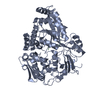
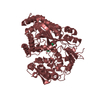
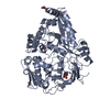
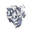
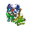
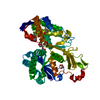
| Title | The structure of the oligopeptide-binding protein, AppA, from Bacillus subtilis in complex with a nonapeptide. | ||||||
 Components Components |
| ||||||
 Keywords Keywords | TRANSPORT PROTEIN / oligopeptide / AppA / transport / Bacillus subtilis | ||||||
| Function / homology |  Function and homology information Function and homology informationestablishment of competence for transformation / peptide transport / peptide transmembrane transporter activity / sporulation resulting in formation of a cellular spore / ATP-binding cassette (ABC) transporter complex / protein transport / periplasmic space Similarity search - Function | ||||||
| Biological species |  | ||||||
| Method |  X-RAY DIFFRACTION / SYNCHROTRON / MOLECULAR REPLACEMENT, MIR / Resolution: 1.55 Å X-RAY DIFFRACTION / SYNCHROTRON / MOLECULAR REPLACEMENT, MIR / Resolution: 1.55 Å | ||||||
 Authors Authors | Levdikov, V.M. / Blagova, E.V. / Brannigan, J.A. / Wright, L. / Vagin, A.A. / Wilkinson, A.J. | ||||||
 Citation Citation | Journal: J.Mol.Biol. / Year: 2005 Title: The structure of the oligopeptide-binding protein, AppA, from Bacillus subtilis in complex with a nonapeptide. Authors: Levdikov, V.M. / Blagova, E.V. / Brannigan, J.A. / Wright, L. / Vagin, A.A. / Wilkinson, A.J. #1: Journal: Acta Crystallogr.,Sect.D / Year: 2004Title: Crystallization of the oligopeptide-binding protein AppA from Bacillus subtilis. Authors: Wright, L. / Blagova, E. / Levdikov, V.M. / Brannigan, J.A. / Pattenden, R.J. / Chambers, J. / Wilkinson, A.J. | ||||||
| History |
|
- Structure visualization
| Structure viewer | Molecule: MolmilJmol/JSmol |
|---|
- Downloads & links
-Download
| PDBx/mmCIF format | 1xoc.cif.gz | 129.5 KB | Display | PDBx/mmCIF format |
|---|---|---|---|---|
| PDB format | pdb1xoc.ent.gz | 98.5 KB | Display | PDB format |
| PDBx/mmJSON format | 1xoc.json.gz | Tree view | PDBx/mmJSON format | |
| Others |  Other downloads Other downloads |
-Validation report
| Arichive directory | https://data.pdbj.org/pub/pdb/validation_reports/xo/1xocftp://data.pdbj.org/pub/pdb/validation_reports/xo/1xoc | HTTPS FTP |
|---|
-Related structure data
| Related structure data |  1dpeS S: Starting model for refinement |
|---|---|
| Similar structure data |


-Links
 PDBj
PDBj


- Assembly
| Deposited unit | 
| ||||||||
|---|---|---|---|---|---|---|---|---|---|
| 1 |
| ||||||||
| 2 | 
| ||||||||
| 3 | 
| ||||||||
| Unit cell |
|
-Components
| #1: Protein | Mass: 59400.691 Da / Num. of mol.: 1 Source method: isolated from a genetically manipulated source Source: (gene. exp.) | ||
|---|---|---|---|
| #2: Protein/peptide | Mass: 1024.063 Da / Num. of mol.: 1 / Source method: obtained synthetically | ||
| #3: Chemical | ChemComp-ZN /   Mass: 65.409 Da / Num. of mol.: 15 / Source method: obtained synthetically / Formula: Zn Mass: 65.409 Da / Num. of mol.: 15 / Source method: obtained synthetically / Formula: Zn#4: Water | ChemComp-HOH / |  Mass: 18.015 Da / Num. of mol.: 441 / Source method: isolated from a natural source / Formula: H2O Mass: 18.015 Da / Num. of mol.: 441 / Source method: isolated from a natural source / Formula: H2O |
-Experimental details
-Experiment
| Experiment | Method: X-RAY DIFFRACTION / Number of used crystals: 1 |
|---|
- Sample preparation
| Crystal | Density Matthews: 2.6 Å3/Da / Density % sol: 52.2 % |
|---|---|
| Crystal grow | Temperature: 291 K / Method: vapor diffusion, hanging drop / pH: 6.5 Details: PEG8000, zinc acetate, MES, pH 6.5, VAPOR DIFFUSION, HANGING DROP, temperature 291K |
-Data collection
| Diffraction | Mean temperature: 100 K |
|---|---|
| Diffraction source | Source: SYNCHROTRON / Site: SRS  / Beamline: PX14.2 / Wavelength: 0.9792 Å / Beamline: PX14.2 / Wavelength: 0.9792 Å |
| Detector | Type: ADSC QUANTUM 4 / Detector: CCD / Date: Sep 18, 2002 / Details: mirrors |
| Radiation | Monochromator: Si 111 CHANNEL / Protocol: SINGLE WAVELENGTH / Monochromatic (M) / Laue (L): M / Scattering type: x-ray |
| Radiation wavelength | Wavelength: 0.9792 Å / Relative weight: 1 |
| Reflection | Resolution: 1.55→50 Å / Num. all: 88121 / Num. obs: 88121 / % possible obs: 97.3 % / Observed criterion σ(F): 0 / Observed criterion σ(I): 0 / Rmerge(I) obs: 0.081 / Net I/σ(I): 18.5 |
| Reflection shell | Resolution: 1.55→1.61 Å / Rmerge(I) obs: 0.788 / Mean I/σ(I) obs: 1.6 / Num. unique all: 6467 / % possible all: 72.6 |
- Processing
| Software |
| ||||||||||||||||||||||||||||||||||||||||||||||||||||||||||||||||||||||||||||||||||||||||||||||||||||||||||||||||||||||||||||||||||||||||||||||||||||||||||||||||
|---|---|---|---|---|---|---|---|---|---|---|---|---|---|---|---|---|---|---|---|---|---|---|---|---|---|---|---|---|---|---|---|---|---|---|---|---|---|---|---|---|---|---|---|---|---|---|---|---|---|---|---|---|---|---|---|---|---|---|---|---|---|---|---|---|---|---|---|---|---|---|---|---|---|---|---|---|---|---|---|---|---|---|---|---|---|---|---|---|---|---|---|---|---|---|---|---|---|---|---|---|---|---|---|---|---|---|---|---|---|---|---|---|---|---|---|---|---|---|---|---|---|---|---|---|---|---|---|---|---|---|---|---|---|---|---|---|---|---|---|---|---|---|---|---|---|---|---|---|---|---|---|---|---|---|---|---|---|---|---|---|---|
| Refinement | Method to determine structure: MOLECULAR REPLACEMENT, MIR Starting model: pdb entry 1DPE Resolution: 1.55→30.57 Å / Cor.coef. Fo:Fc: 0.97 / Cor.coef. Fo:Fc free: 0.959 / SU B: 3.343 / SU ML: 0.061 / Isotropic thermal model: Isotropic / Cross valid method: THROUGHOUT / σ(F): 0 / σ(I): 0 / ESU R: 0.074 / ESU R Free: 0.076 / Stereochemistry target values: MAXIMUM LIKELIHOOD / Details: HYDROGENS HAVE BEEN ADDED IN THE RIDING POSITIONS
| ||||||||||||||||||||||||||||||||||||||||||||||||||||||||||||||||||||||||||||||||||||||||||||||||||||||||||||||||||||||||||||||||||||||||||||||||||||||||||||||||
| Solvent computation | Ion probe radii: 0.8 Å / Shrinkage radii: 0.8 Å / VDW probe radii: 1.2 Å / Solvent model: MASK | ||||||||||||||||||||||||||||||||||||||||||||||||||||||||||||||||||||||||||||||||||||||||||||||||||||||||||||||||||||||||||||||||||||||||||||||||||||||||||||||||
| Displacement parameters | Biso mean: 27.752 Å2
| ||||||||||||||||||||||||||||||||||||||||||||||||||||||||||||||||||||||||||||||||||||||||||||||||||||||||||||||||||||||||||||||||||||||||||||||||||||||||||||||||
| Refinement step | Cycle: LAST / Resolution: 1.55→30.57 Å
| ||||||||||||||||||||||||||||||||||||||||||||||||||||||||||||||||||||||||||||||||||||||||||||||||||||||||||||||||||||||||||||||||||||||||||||||||||||||||||||||||
| Refine LS restraints |
| ||||||||||||||||||||||||||||||||||||||||||||||||||||||||||||||||||||||||||||||||||||||||||||||||||||||||||||||||||||||||||||||||||||||||||||||||||||||||||||||||
| LS refinement shell | Resolution: 1.55→1.587 Å / Total num. of bins used: 20
|
