 Movie
Movie Controller

[English] 日本語
 Yorodumi
- PDB-3fh6: Crystal structure of the resting state maltose transporter from E... -
+ Open data

- Basic information
| Entry | Database: PDB / ID: 3fh6 | ||||||
|---|---|---|---|---|---|---|---|
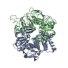
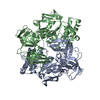
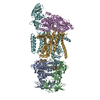
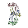
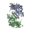
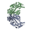
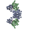
| Title | Crystal structure of the resting state maltose transporter from E. coli | ||||||
 Components Components |
| ||||||
 Keywords Keywords | TRANSPORT PROTEIN / maltose transporter / ground state / ABC transporter / membrane protein / Cell inner membrane / Cell membrane / Membrane / Sugar transport / Transmembrane / Transport / ATP-binding / Hydrolase / Nucleotide-binding | ||||||
| Function / homology |  Function and homology information Function and homology informationABC-type maltose transporter / ABC-type maltose transporter activity / negative regulation of maltose transport / enzyme IIA-maltose transporter complex / negative regulation of transmembrane transport / maltose transport complex / maltose transport / maltodextrin transmembrane transport / ATP-binding cassette (ABC) transporter complex, substrate-binding subunit-containing / ATP-binding cassette (ABC) transporter complex ...ABC-type maltose transporter / ABC-type maltose transporter activity / negative regulation of maltose transport / enzyme IIA-maltose transporter complex / negative regulation of transmembrane transport / maltose transport complex / maltose transport / maltodextrin transmembrane transport / ATP-binding cassette (ABC) transporter complex, substrate-binding subunit-containing / ATP-binding cassette (ABC) transporter complex / DNA-binding transcription factor binding / DNA damage response / ATP hydrolysis activity / ATP binding / membrane / plasma membrane Similarity search - Function | ||||||
| Biological species |  | ||||||
| Method |  X-RAY DIFFRACTION / SYNCHROTRON / MAD / Resolution: 4.5 Å X-RAY DIFFRACTION / SYNCHROTRON / MAD / Resolution: 4.5 Å | ||||||
 Authors Authors | Khare, D. / Oldham, M.L. / Orelle, C. / Davidson, A.L. / Chen, J. | ||||||
 Citation Citation | Journal: Mol.Cell / Year: 2009 Title: Alternating access in maltose transporter mediated by rigid-body rotations. Authors: Khare, D. / Oldham, M.L. / Orelle, C. / Davidson, A.L. / Chen, J. | ||||||
| History |
|
- Structure visualization
| Structure viewer | Molecule: MolmilJmol/JSmol |
|---|
- Downloads & links
-Download
| PDBx/mmCIF format | 3fh6.cif.gz | 473.4 KB | Display | PDBx/mmCIF format |
|---|---|---|---|---|
| PDB format | pdb3fh6.ent.gz | 383.5 KB | Display | PDB format |
| PDBx/mmJSON format | 3fh6.json.gz | Tree view | PDBx/mmJSON format | |
| Others |  Other downloads Other downloads |
-Validation report
| Summary document | 3fh6_validation.pdf.gz | 526.4 KB | Display | wwPDB validaton report |
|---|---|---|---|---|
| Full document | 3fh6_full_validation.pdf.gz | 885.6 KB | Display | |
| Data in XML | 3fh6_validation.xml.gz | 141.1 KB | Display | |
| Data in CIF | 3fh6_validation.cif.gz | 185.1 KB | Display | |
| Arichive directory | https://data.pdbj.org/pub/pdb/validation_reports/fh/3fh6ftp://data.pdbj.org/pub/pdb/validation_reports/fh/3fh6 | HTTPS FTP |
-Related structure data


-Links
 PDBj
PDBj

- Assembly
| Deposited unit | 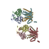
| ||||||||
|---|---|---|---|---|---|---|---|---|---|
| 1 | 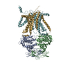
| ||||||||
| 2 | 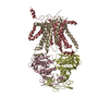
| ||||||||
| Unit cell |
| ||||||||
| Noncrystallographic symmetry (NCS) | NCS oper: (Code: given Matrix: (0.88774, 0.08265, 0.45286), Vector: |
-Components
| #1: Protein | Mass: 53093.113 Da / Num. of mol.: 2 Source method: isolated from a genetically manipulated source Source: (gene. exp.) #2: Protein | Mass: 32246.227 Da / Num. of mol.: 2 Source method: isolated from a genetically manipulated source Source: (gene. exp.) #3: Protein | Mass: 42184.535 Da / Num. of mol.: 4 Source method: isolated from a genetically manipulated source Source: (gene. exp.) |
|---|
-Experimental details
-Experiment
| Experiment | Method: X-RAY DIFFRACTION / Number of used crystals: 1 |
|---|
- Sample preparation
| Crystal | Density Matthews: 5.79 Å3/Da / Density % sol: 78.8 % |
|---|---|
| Crystal grow | Temperature: 293 K / Method: vapor diffusion, sitting drop / pH: 6.5 Details: 11.5 % PEG 4000, 0.1M ADA pH 6.5, 0.1 M NaCl, 0.1 M Li2SO4, VAPOR DIFFUSION, SITTING DROP, temperature 293K |
-Data collection
| Diffraction | Mean temperature: 100 K | |||||||||||||||||||||||||||||||||||||||||||||||||||||||||||||||||||||||||||||
|---|---|---|---|---|---|---|---|---|---|---|---|---|---|---|---|---|---|---|---|---|---|---|---|---|---|---|---|---|---|---|---|---|---|---|---|---|---|---|---|---|---|---|---|---|---|---|---|---|---|---|---|---|---|---|---|---|---|---|---|---|---|---|---|---|---|---|---|---|---|---|---|---|---|---|---|---|---|---|
| Diffraction source | Source: SYNCHROTRON / Site: APS  / Beamline: 23-ID-D / Wavelength: 0.97942, 0.9794, 0.9795, 0.9494 / Beamline: 23-ID-D / Wavelength: 0.97942, 0.9794, 0.9795, 0.9494 | |||||||||||||||||||||||||||||||||||||||||||||||||||||||||||||||||||||||||||||
| Detector | Type: MARMOSAIC 300 mm CCD / Detector: CCD / Date: Dec 10, 2007 | |||||||||||||||||||||||||||||||||||||||||||||||||||||||||||||||||||||||||||||
| Radiation | Monochromator: Si(111) double crystal / Protocol: MAD / Scattering type: x-ray | |||||||||||||||||||||||||||||||||||||||||||||||||||||||||||||||||||||||||||||
| Radiation wavelength |
| |||||||||||||||||||||||||||||||||||||||||||||||||||||||||||||||||||||||||||||
| Reflection | Resolution: 4.5→100 Å / Num. obs: 40160 / % possible obs: 87.5 % / Redundancy: 3.4 % / Rmerge(I) obs: 0.126 / Χ2: 1.975 / Net I/σ(I): 11.126 | |||||||||||||||||||||||||||||||||||||||||||||||||||||||||||||||||||||||||||||
| Reflection shell |
|
-Phasing
| Phasing | Method: MAD |
|---|
- Processing
| Software |
| ||||||||||||||||||||||||||||||||||||
|---|---|---|---|---|---|---|---|---|---|---|---|---|---|---|---|---|---|---|---|---|---|---|---|---|---|---|---|---|---|---|---|---|---|---|---|---|---|
| Refinement | Method to determine structure: MAD Starting model: PDB entries 1Q1B, 1Q1E, 2R6G Resolution: 4.5→50 Å / Occupancy max: 1 / Occupancy min: 1 / σ(F): 0 Details: The structure was refined with strict NCS restraints applied to the refined molecules F,G,A,B. The molecules H,I,C,D that build the complete asymmetric unit, are the respective NCS-mates
| ||||||||||||||||||||||||||||||||||||
| Solvent computation | Bsol: 137.42 Å2 | ||||||||||||||||||||||||||||||||||||
| Displacement parameters | Biso max: 521.43 Å2 / Biso mean: 300.466 Å2 / Biso min: 94.21 Å2 | ||||||||||||||||||||||||||||||||||||
| Refinement step | Cycle: LAST / Resolution: 4.5→50 Å
| ||||||||||||||||||||||||||||||||||||
| Refine LS restraints |
| ||||||||||||||||||||||||||||||||||||
| Xplor file | Serial no: 1 / Param file: protein_between.param |
