[English] 日本語
Yorodumi
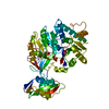
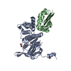
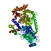
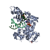
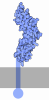
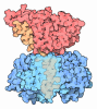
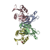
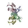
- PDB-3ff7: Structure of NK cell receptor KLRG1 bound to E-cadherin -

+
Open data


ID or keywords:

Loading...

-
Basic information

Entry
Database: PDB / ID: 3ff7
TitleStructure of NK cell receptor KLRG1 bound to E-cadherin
Components
  • Epithelial cadherin
  • Killer cell lectin-like receptor subfamily G member 1
KeywordsCell adhesion/Immune system / KLRG1-cadherin complex / Calcium / Cell adhesion / Cell junction / Cell membrane / Cleavage on pair of basic residues / Disease mutation / Glycoprotein / Membrane / Phosphoprotein / Polymorphism / Transmembrane / Alternative splicing / Lectin / Receptor / Signal-anchor / Cell adhesion-Immune system COMPLEX
Function / homology
Function and homology information


response to heparin / desmosome assembly / pituitary gland development / response to Gram-positive bacterium / gamma-catenin binding / desmosome / Regulation of MITF-M-dependent genes involved in extracellular matrix, focal adhesion and epithelial-to-mesenchymal transition / negative regulation of axon extension / Regulation of CDH1 mRNA translation by microRNAs / cellular response to lithium ion ...response to heparin / desmosome assembly / pituitary gland development / response to Gram-positive bacterium / gamma-catenin binding / desmosome / Regulation of MITF-M-dependent genes involved in extracellular matrix, focal adhesion and epithelial-to-mesenchymal transition / negative regulation of axon extension / Regulation of CDH1 mRNA translation by microRNAs / cellular response to lithium ion / cellular response to indole-3-methanol / cell adhesion mediator activity / calcium-dependent cell-cell adhesion / adherens junction organization / cell-cell adhesion mediated by cadherin / regulation of protein catabolic process at postsynapse, modulating synaptic transmission / flotillin complex / Developmental Lineage of Mammary Stem Cells / Formation of definitive endoderm / Apoptotic cleavage of cell adhesion proteins / catenin complex / Regulation of CDH1 Function / cell-cell junction assembly / Adherens junctions interactions / negative regulation of cell-cell adhesion / GTPase activating protein binding / ankyrin binding / Regulation of CDH1 posttranslational processing and trafficking to plasma membrane / apical junction complex / homophilic cell-cell adhesion / Developmental Lineage of Mammary Gland Myoepithelial Cells / Developmental Lineage of Mammary Gland Luminal Epithelial Cells / lateral plasma membrane / Integrin cell surface interactions / cellular defense response / cell morphogenesis / RHO GTPases activate IQGAPs / Degradation of the extracellular matrix / positive regulation of protein localization / synapse assembly / Transcriptional and post-translational regulation of MITF-M expression and activity / cell adhesion molecule binding / negative regulation of cell migration / protein tyrosine kinase binding / protein localization to plasma membrane / InlA-mediated entry of Listeria monocytogenes into host cells / adherens junction / positive regulation of protein import into nucleus / trans-Golgi network / cell-cell adhesion / response to toxic substance / cell junction / Golgi lumen / beta-catenin binding / Degradation of CDH1 / neuron projection development / Immunoregulatory interactions between a Lymphoid and a non-Lymphoid cell / cytoplasmic side of plasma membrane / actin cytoskeleton / regulation of gene expression / cell migration / lamellipodium / carbohydrate binding / signaling receptor activity / early endosome membrane / cell surface receptor signaling pathway / postsynapse / response to xenobiotic stimulus / inflammatory response / cadherin binding / endoplasmic reticulum lumen / Golgi membrane / innate immune response / lysosomal membrane / calcium ion binding / positive regulation of DNA-templated transcription / endoplasmic reticulum membrane / perinuclear region of cytoplasm / glutamatergic synapse / Golgi apparatus / extracellular exosome / extracellular region / membrane / identical protein binding / plasma membrane / cytoplasm
Similarity search - Function
Killer cell lectin-like receptor subfamily G member 1 / Cadherin prodomain like / Cadherin prodomain / Cadherin prodomain like / Natural killer cell receptor-like, C-type lectin-like domain / Cadherin, Y-type LIR-motif / Cadherin, Y-type LIR-motif / Cadherin / Catenin binding domain superfamily / Cadherins ...Killer cell lectin-like receptor subfamily G member 1 / Cadherin prodomain like / Cadherin prodomain / Cadherin prodomain like / Natural killer cell receptor-like, C-type lectin-like domain / Cadherin, Y-type LIR-motif / Cadherin, Y-type LIR-motif / Cadherin / Catenin binding domain superfamily / Cadherins / Cadherin conserved site / Cadherin domain signature. / Cadherin repeats. / Cadherin domain / Cadherin-like / Cadherins domain profile. / Cadherin-like superfamily / Mannose-Binding Protein A; Chain A / Mannose-Binding Protein A, subunit A / C-type lectin domain profile. / C-type lectin-like / C-type lectin (CTL) or carbohydrate-recognition domain (CRD) / Lectin C-type domain / C-type lectin-like/link domain superfamily / C-type lectin fold / Roll / Immunoglobulin-like / Sandwich / Mainly Beta / Alpha Beta
Similarity search - Domain/homology
ACETIC ACID / Cadherin-1 / Killer cell lectin-like receptor subfamily G member 1
Similarity search - Component
Biological speciesHomo sapiens (human)
MethodX-RAY DIFFRACTION / SYNCHROTRON / MOLECULAR REPLACEMENT / molecular replacement / Resolution: 1.8 Å
AuthorsLi, Y. / Mariuzza, R.A.
CitationJournal: Immunity / Year: 2009
Title: Structure of natural killer cell receptor KLRG1 bound to E-cadherin reveals basis for MHC-independent missing self recognition.
Authors: Li, Y. / Hofmann, M. / Wang, Q. / Teng, L. / Chlewicki, L.K. / Pircher, H. / Mariuzza, R.A.
History
DepositionDec 2, 2008Deposition site: RCSB / Processing site: RCSB
Revision 1.0Jul 28, 2009Provider: repository / Type: Initial release
Revision 1.1Jul 13, 2011Group: Version format compliance
Revision 1.2Nov 1, 2017Group: Advisory / Refinement description / Category: pdbx_unobs_or_zero_occ_atoms / software
Revision 1.3Oct 20, 2021Group: Advisory / Database references / Derived calculations
Category: database_2 / pdbx_unobs_or_zero_occ_atoms ...database_2 / pdbx_unobs_or_zero_occ_atoms / struct_ref_seq_dif / struct_site
Item: _database_2.pdbx_DOI / _database_2.pdbx_database_accession ..._database_2.pdbx_DOI / _database_2.pdbx_database_accession / _struct_ref_seq_dif.details / _struct_site.pdbx_auth_asym_id / _struct_site.pdbx_auth_comp_id / _struct_site.pdbx_auth_seq_id
Revision 1.4Dec 27, 2023Group: Data collection / Category: chem_comp_atom / chem_comp_bond
Revision 1.5Apr 3, 2024Group: Refinement description / Category: pdbx_initial_refinement_model
Revision 1.6Oct 16, 2024Group: Structure summary / Category: pdbx_entry_details / pdbx_modification_feature

-
Structure visualization

Structure viewerMolecule:
MolmilJmol/JSmol

Downloads & links

-
Assembly

Deposited unit
A: Epithelial cadherin
B: Epithelial cadherin
C: Killer cell lectin-like receptor subfamily G member 1
D: Killer cell lectin-like receptor subfamily G member 1
hetero molecules


Theoretical massNumber of molelcules
Total (without water)48,2545
Polymers48,1944
Non-polymers601
Water5,567309
1


  • Idetical with deposited unit
  • defined by author
TypeNameSymmetry operationNumber
identity operation1_555x,y,z1
Unit cell
Length a, b, c (Å)54.120, 83.411, 56.508
Angle α, β, γ (deg.)90.000, 106.100, 90.000
Int Tables number4
Space group name H-MP1211

-
Components

#1: Protein Epithelial cadherin / E-cadherin / Cadherin-1 / Uvomorulin / CAM 120/80 / E-Cad/CTF1 / E-Cad/CTF2 / E-Cad/CTF3


Mass: 11124.637 Da / Num. of mol.: 2 / Fragment: UNP residues 155-253, Cadherin 1 domain / Mutation: C9L
Source method: isolated from a genetically manipulated source
Source: (gene. exp.) Homo sapiens (human) / Gene: CDH1, CDHE, UVO / Production host: Escherichia coli (E. coli) / References: UniProt: P12830
#2: Protein Killer cell lectin-like receptor subfamily G member 1 / Mast cell function-associated antigen / MAFA-like receptor / ITIM-containing receptor MAFA-L / C- ...Mast cell function-associated antigen / MAFA-like receptor / ITIM-containing receptor MAFA-L / C-type lectin domain family 15 member A


Mass: 12972.562 Da / Num. of mol.: 2 / Fragment: UNP residues 75-186, C-type lectin domain / Mutation: C131S
Source method: isolated from a genetically manipulated source
Source: (gene. exp.) Homo sapiens (human) / Gene: KLRG1, CLEC15A, MAFA, MAFAL / Production host: Escherichia coli (E. coli) / References: UniProt: Q96E93
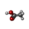
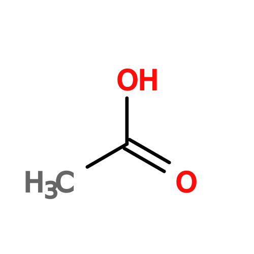
#3: Chemical ChemComp-ACY / ACETIC ACID


Mass: 60.052 Da / Num. of mol.: 1 / Source method: obtained synthetically / Formula: C2H4O2

#4: Water ChemComp-HOH / water


Mass: 18.015 Da / Num. of mol.: 309 / Source method: isolated from a natural source / Formula: H2O
Has protein modificationY

-
Experimental details

-
Experiment

ExperimentMethod: X-RAY DIFFRACTION / Number of used crystals: 1

-
Sample preparation

CrystalDensity Matthews: 2.54 Å3/Da / Density % sol: 51.63 %
Crystal growTemperature: 298 K / Method: vapor diffusion / pH: 4.6 / Details: PEG4000, pH 4.6, vapor diffusion, temperature 298K

-
Data collection

DiffractionMean temperature: 100 K
Diffraction sourceSource: SYNCHROTRON / Site: NSLS / Beamline: X29A / Wavelength: 1.502 Å
DetectorType: ADSC QUANTUM 315 / Detector: CCD / Date: Jul 7, 2008
RadiationProtocol: SINGLE WAVELENGTH / Scattering type: x-ray
Radiation wavelengthWavelength: 1.502 Å / Relative weight: 1
ReflectionResolution: 1.65→30 Å / Num. obs: 57098 / % possible obs: 98.6 % / Redundancy: 7.3 % / Rmerge(I) obs: 0.064 / Χ2: 1.289 / Net I/σ(I): 21.542
Reflection shell
Resolution (Å)Redundancy (%)Rmerge(I) obsNum. unique allΧ2% possible all
1.65-1.714.90.56351041.00288.3
1.71-1.786.50.44356791.02798.6
1.78-1.867.30.31557381.042100
1.86-1.967.40.20658071.142100
1.96-2.087.50.12157781.206100
2.08-2.247.50.08957771.222100
2.24-2.467.40.0757961.189100
2.46-2.827.50.05657831.115100
2.82-3.557.40.05258201.14499.9
3.55-309.60.05858162.28398.8

-
Phasing

PhasingMethod: molecular replacement
Phasing MRModel details: Phaser MODE: MR_AUTO
Highest resolutionLowest resolution
Rotation2.5 Å27.71 Å
Translation2.5 Å27.71 Å

-
Processing

Software
NameVersionClassificationNB
DENZOdata reduction
SCALEPACKdata scaling
PHASERphasing
CNSrefinement
PDB_EXTRACT3.006data extraction
ADSCQuantumdata collection
RefinementMethod to determine structure: MOLECULAR REPLACEMENT
Starting model: PDB ENTRY 3FF7
Resolution: 1.8→30 Å / Occupancy max: 1 / Occupancy min: 0 / σ(F): 0
RfactorNum. reflection% reflection
Rfree0.246 1340 3 %
Rwork0.214 --
obs-44610 99.8 %
Solvent computationBsol: 35.739 Å2
Displacement parametersBiso max: 64.81 Å2 / Biso mean: 28.672 Å2 / Biso min: 11.36 Å2
Baniso -1Baniso -2Baniso -3
1--0.125 Å20 Å2-0.121 Å2
2--3.401 Å20 Å2
3----3.276 Å2
Refinement stepCycle: LAST / Resolution: 1.8→30 Å
ProteinNucleic acidLigandSolventTotal
Num. atoms3335 0 0 313 3648
Refine LS restraints
Refine-IDTypeDev ideal
X-RAY DIFFRACTIONc_bond_d0.009
X-RAY DIFFRACTIONc_angle_d1.559
Xplor file
Refine-IDSerial noParam file
X-RAY DIFFRACTION1CNS_TOPPAR:protein_rep.param
X-RAY DIFFRACTION2CNS_TOPPAR:water.param
X-RAY DIFFRACTION3acy.param

+
About Yorodumi

-
News

-
Feb 9, 2022. New format data for meta-information of EMDB entries

New format data for meta-information of EMDB entries

  • Version 3 of the EMDB header file is now the official format.
  • The previous official version 1.9 will be removed from the archive.

Related info.:EMDB header

External links:wwPDB to switch to version 3 of the EMDB data model

-
Aug 12, 2020. Covid-19 info

Covid-19 info

URL: https://pdbj.org/emnavi/covid19.php

New page: Covid-19 featured information page in EM Navigator.

Related info.:Covid-19 info / Mar 5, 2020. Novel coronavirus structure data

+
Mar 5, 2020. Novel coronavirus structure data

Novel coronavirus structure data

Related info.:Yorodumi Speices / Aug 12, 2020. Covid-19 info

External links:COVID-19 featured content - PDBj / Molecule of the Month (242):Coronavirus Proteases

+
Jan 31, 2019. EMDB accession codes are about to change! (news from PDBe EMDB page)

EMDB accession codes are about to change! (news from PDBe EMDB page)

  • The allocation of 4 digits for EMDB accession codes will soon come to an end. Whilst these codes will remain in use, new EMDB accession codes will include an additional digit and will expand incrementally as the available range of codes is exhausted. The current 4-digit format prefixed with “EMD-” (i.e. EMD-XXXX) will advance to a 5-digit format (i.e. EMD-XXXXX), and so on. It is currently estimated that the 4-digit codes will be depleted around Spring 2019, at which point the 5-digit format will come into force.
  • The EM Navigator/Yorodumi systems omit the EMD- prefix.

Related info.:Q: What is EMD? / ID/Accession-code notation in Yorodumi/EM Navigator

External links:EMDB Accession Codes are Changing Soon! / Contact to PDBj

+
Jul 12, 2017. Major update of PDB

Major update of PDB

  • wwPDB released updated PDB data conforming to the new PDBx/mmCIF dictionary.
  • This is a major update changing the version number from 4 to 5, and with Remediation, in which all the entries are updated.
  • In this update, many items about electron microscopy experimental information are reorganized (e.g. em_software).
  • Now, EM Navigator and Yorodumi are based on the updated data.

External links:wwPDB Remediation / Enriched Model Files Conforming to OneDep Data Standards Now Available in the PDB FTP Archive

-
Yorodumi

Thousand views of thousand structures

  • Yorodumi is a browser for structure data from EMDB, PDB, SASBDB, etc.
  • This page is also the successor to EM Navigator detail page, and also detail information page/front-end page for Omokage search.
  • The word "yorodu" (or yorozu) is an old Japanese word meaning "ten thousand". "mi" (miru) is to see.

Related info.:EMDB / PDB / SASBDB / Comparison of 3 databanks / Yorodumi Search / Aug 31, 2016. New EM Navigator & Yorodumi / Yorodumi Papers / Jmol/JSmol / Function and homology information / Changes in new EM Navigator and Yorodumi

Read more