 Movie
Movie Controller


+ Open data

- Basic information
| Entry | Database: PDB / ID: 5d1u | ||||||
|---|---|---|---|---|---|---|---|
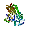
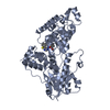
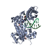
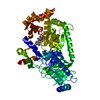
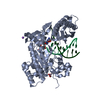
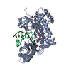
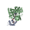
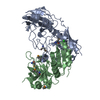
| Title | Anthrax toxin lethal factor with hydroxamic acid inhibitor | ||||||
 Components Components | Lethal factor | ||||||
 Keywords Keywords | Hydrolase/hydrolase inhibitor / Anthrax toxin / lethal factor / metalloproteinase / metalloprotease / structural dynamics / ligand-induced conformational change / Hydrolase-hydrolase inhibitor complex | ||||||
| Function / homology |  Function and homology information Function and homology informationanthrax lethal factor endopeptidase / host cell cytosol / Uptake and function of anthrax toxins / metalloendopeptidase activity / metallopeptidase activity / toxin activity / proteolysis / extracellular region / zinc ion binding Similarity search - Function | ||||||
| Biological species |  | ||||||
| Method |  X-RAY DIFFRACTION / SYNCHROTRON / MOLECULAR REPLACEMENT / Resolution: 2.8503 Å X-RAY DIFFRACTION / SYNCHROTRON / MOLECULAR REPLACEMENT / Resolution: 2.8503 Å | ||||||
 Authors Authors | Maize, K.M. / Finzel, B.C. | ||||||
| Funding support |  United States, 1items United States, 1items
| ||||||
 Citation Citation | Journal: J.Med.Chem. / Year: 2015 Title: Probing the S2' Subsite of the Anthrax Toxin Lethal Factor Using Novel N-Alkylated Hydroxamates. Authors: Kurbanov, E.K. / Chiu, T.L. / Solberg, J. / Francis, S. / Maize, K.M. / Fernandez, J. / Johnson, R.L. / Hawkinson, J.E. / Walters, M.A. / Finzel, B.C. / Amin, E.A. | ||||||
| History |
|
- Structure visualization
| Structure viewer | Molecule: MolmilJmol/JSmol |
|---|
- Downloads & links
-Download
| PDBx/mmCIF format | 5d1u.cif.gz | 113.2 KB | Display | PDBx/mmCIF format |
|---|---|---|---|---|
| PDB format | pdb5d1u.ent.gz | 82.8 KB | Display | PDB format |
| PDBx/mmJSON format | 5d1u.json.gz | Tree view | PDBx/mmJSON format | |
| Others |  Other downloads Other downloads |
-Validation report
| Arichive directory | https://data.pdbj.org/pub/pdb/validation_reports/d1/5d1uftp://data.pdbj.org/pub/pdb/validation_reports/d1/5d1u | HTTPS FTP |
|---|
-Related structure data
| Related structure data |  4wf6C 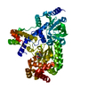 5d1sC  5d1tC  1yqyS S: Starting model for refinement C: citing same article ( |
|---|---|
| Similar structure data |


-Links
 PDBj
PDBj


- Assembly
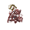
| Deposited unit | 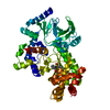
| ||||||||
|---|---|---|---|---|---|---|---|---|---|
| 1 |
| ||||||||
| Unit cell |
|
-Components
| #1: Protein | Mass: 60436.047 Da / Num. of mol.: 1 / Fragment: residues 298-809 / Mutation: A299S Source method: isolated from a genetically manipulated source Source: (gene. exp.) References: UniProt: P15917, anthrax lethal factor endopeptidase |
|---|---|
| #2: Chemical | ChemComp-56P /   Mass: 375.459 Da / Num. of mol.: 1 / Source method: obtained synthetically / Formula: C16H26FN3O4S Mass: 375.459 Da / Num. of mol.: 1 / Source method: obtained synthetically / Formula: C16H26FN3O4S |
| #3: Chemical | ChemComp-ZN /   Mass: 65.409 Da / Num. of mol.: 1 / Source method: obtained synthetically / Formula: Zn Mass: 65.409 Da / Num. of mol.: 1 / Source method: obtained synthetically / Formula: Zn |
| #4: Water | ChemComp-HOH /  Mass: 18.015 Da / Num. of mol.: 17 / Source method: isolated from a natural source / Formula: H2O Mass: 18.015 Da / Num. of mol.: 17 / Source method: isolated from a natural source / Formula: H2O |
-Experimental details
-Experiment
| Experiment | Method: X-RAY DIFFRACTION / Number of used crystals: 1 |
|---|
- Sample preparation
| Crystal | Density Matthews: 2.56 Å3/Da / Density % sol: 52.05 % |
|---|---|
| Crystal grow | Temperature: 286 K / Method: vapor diffusion, hanging drop / pH: 6.8 Details: 11-16% PEG 8K, 50 mM Bis-Tris, 100 mM magnesium acetate |
-Data collection
| Diffraction | Mean temperature: 100 K | |||||||||||||||
|---|---|---|---|---|---|---|---|---|---|---|---|---|---|---|---|---|
| Diffraction source | Source: SYNCHROTRON / Site: APS / Beamline: 17-ID / Wavelength: 1 Å | |||||||||||||||
| Detector | Type: DECTRIS PILATUS 6M / Detector: PIXEL / Date: Dec 1, 2014 | |||||||||||||||
| Radiation | Protocol: SINGLE WAVELENGTH / Monochromatic (M) / Laue (L): M / Scattering type: x-ray | |||||||||||||||
| Radiation wavelength | Wavelength: 1 Å / Relative weight: 1 | |||||||||||||||
| Reflection | Resolution: 2.85→77.293 Å / Num. obs: 15129 / % possible obs: 100 % / Redundancy: 6.4 % / Biso Wilson estimate: 52.08 Å2 / Rmerge(I) obs: 0.063 / Net I/σ(I): 24.1 / Num. measured all: 96780 | |||||||||||||||
| Reflection shell | Diffraction-ID: 1 / Rejects: _ / % possible all: 100
|
- Processing
| Software |
| ||||||||||||||||||||||||||||||||||||
|---|---|---|---|---|---|---|---|---|---|---|---|---|---|---|---|---|---|---|---|---|---|---|---|---|---|---|---|---|---|---|---|---|---|---|---|---|---|
| Refinement | Method to determine structure: MOLECULAR REPLACEMENT Starting model: 1YQY Resolution: 2.8503→39.632 Å / SU ML: 0.38 / Cross valid method: FREE R-VALUE / σ(F): 1.34 / Phase error: 27.24 / Stereochemistry target values: ML
| ||||||||||||||||||||||||||||||||||||
| Solvent computation | Shrinkage radii: 0.9 Å / VDW probe radii: 1.11 Å / Solvent model: FLAT BULK SOLVENT MODEL | ||||||||||||||||||||||||||||||||||||
| Displacement parameters | Biso max: 121.13 Å2 / Biso mean: 51.3988 Å2 / Biso min: 20.71 Å2 | ||||||||||||||||||||||||||||||||||||
| Refinement step | Cycle: final / Resolution: 2.8503→39.632 Å
| ||||||||||||||||||||||||||||||||||||
| Refine LS restraints |
| ||||||||||||||||||||||||||||||||||||
| LS refinement shell | Refine-ID: X-RAY DIFFRACTION / Total num. of bins used: 5 / % reflection obs: 100 %
|
