 Movie
Movie Controller

[English] 日本語
 Yorodumi
- PDB-3fe9: Crystal structure of a pheromone binding protein from Apis mellif... -
+ Open data

- Basic information
| Entry | Database: PDB / ID: 3fe9 | ||||||
|---|---|---|---|---|---|---|---|
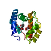
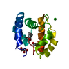
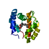
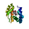
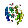
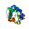
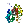
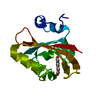
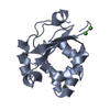
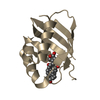
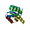
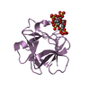
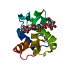
| Title | Crystal structure of a pheromone binding protein from Apis mellifera with a serendipitous ligand soaked at pH 7.0 | ||||||
 Components Components | Pheromone-binding protein ASP1 | ||||||
 Keywords Keywords | Pheromone binding protein / honey bee / Apis mellifera / signal transduction / queen mandibular protein | ||||||
| Function / homology |  Function and homology information Function and homology information | ||||||
| Biological species |  | ||||||
| Method |  X-RAY DIFFRACTION / SYNCHROTRON / FOURIER SYNTHESIS / Resolution: 1.75 Å X-RAY DIFFRACTION / SYNCHROTRON / FOURIER SYNTHESIS / Resolution: 1.75 Å | ||||||
 Authors Authors | Pesenti, M.E. / Spinelli, S. / Bezirard, V. / Briand, L. / Pernollet, J.C. / Tegoni, M. / Cambillau, C. | ||||||
 Citation Citation | Journal: To be Published Title: Queen bee pheromone binding protein pH induced domain-swapping favors pheromone release Authors: Pesenti, M.E. / Spinelli, S. / Bezirard, V. / Briand, L. / Pernollet, J.C. / Tegoni, M. / Cambillau, C. #1: Journal: J.Mol.Biol. / Year: 2008Title: Structural basis of the honey bee PBP pheromone and pH-induced conformational change Authors: Pesenti, M.E. / Spinelli, S. / Bezirard, V. / Briand, L. / Pernollet, J.C. / Tegoni, M. / Cambillau, C. | ||||||
| History |
|
- Structure visualization
| Structure viewer | Molecule: MolmilJmol/JSmol |
|---|
- Downloads & links
-Download
| PDBx/mmCIF format | 3fe9.cif.gz | 42.8 KB | Display | PDBx/mmCIF format |
|---|---|---|---|---|
| PDB format | pdb3fe9.ent.gz | 28.7 KB | Display | PDB format |
| PDBx/mmJSON format | 3fe9.json.gz | Tree view | PDBx/mmJSON format | |
| Others |  Other downloads Other downloads |
-Validation report
| Arichive directory | https://data.pdbj.org/pub/pdb/validation_reports/fe/3fe9ftp://data.pdbj.org/pub/pdb/validation_reports/fe/3fe9 | HTTPS FTP |
|---|
-Related structure data
| Related structure data | 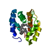 3fe6C  3fe8C 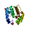 2h8vS S: Starting model for refinement C: citing same article ( |
|---|---|
| Similar structure data |


-Links
 PDBj
PDBj- Assembly
| Deposited unit | 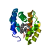
| |||||||||||||||
|---|---|---|---|---|---|---|---|---|---|---|---|---|---|---|---|---|
| 1 |
| |||||||||||||||
| Unit cell |
| |||||||||||||||
| Components on special symmetry positions |
|
-Components
| #1: Protein | Mass: 13194.789 Da / Num. of mol.: 1 / Fragment: UNP residues 26-144 Source method: isolated from a genetically manipulated source Source: (gene. exp.)  Pichia pastoris (fungus) / References: UniProt: Q9U9J6 Pichia pastoris (fungus) / References: UniProt: Q9U9J6 |
|---|---|
| #2: Chemical | ChemComp-CMJ / (  Mass: 605.159 Da / Num. of mol.: 1 / Source method: obtained synthetically / Formula: C43H88 Mass: 605.159 Da / Num. of mol.: 1 / Source method: obtained synthetically / Formula: C43H88 |
| #3: Chemical | ChemComp-CL /   Mass: 35.453 Da / Num. of mol.: 1 / Source method: obtained synthetically / Formula: Cl Mass: 35.453 Da / Num. of mol.: 1 / Source method: obtained synthetically / Formula: Cl |
| #4: Water | ChemComp-HOH /  Mass: 18.015 Da / Num. of mol.: 159 / Source method: isolated from a natural source / Formula: H2O Mass: 18.015 Da / Num. of mol.: 159 / Source method: isolated from a natural source / Formula: H2O |
| Has protein modification | Y |
-Experimental details
-Experiment
| Experiment | Method: X-RAY DIFFRACTION / Number of used crystals: 1 |
|---|
- Sample preparation
| Crystal | Density Matthews: 3.1 Å3/Da / Density % sol: 60.31 % |
|---|---|
| Crystal grow | Temperature: 293 K / Method: vapor diffusion, sitting drop / pH: 5.5 Details: 1.7M ammonium sulfate, 0.1M sodium citrate, pH5.5, VAPOR DIFFUSION, SITTING DROP, temperature 293K |
-Data collection
| Diffraction | Mean temperature: 100 K |
|---|---|
| Diffraction source | Source: SYNCHROTRON / Site: ESRF  / Beamline: ID14-2 / Wavelength: 0.933 Å / Beamline: ID14-2 / Wavelength: 0.933 Å |
| Detector | Type: ADSC QUANTUM 4 / Detector: CCD / Date: Nov 24, 2007 / Details: tiroidal mirror |
| Radiation | Monochromator: Si 111 / Protocol: SINGLE WAVELENGTH / Monochromatic (M) / Laue (L): M / Scattering type: x-ray |
| Radiation wavelength | Wavelength: 0.933 Å / Relative weight: 1 |
| Reflection | Resolution: 1.75→41.96 Å / Num. all: 16926 / Num. obs: 16926 / % possible obs: 99.9 % / Redundancy: 7.9 % / Biso Wilson estimate: 21.99 Å2 / Rmerge(I) obs: 0.087 / Rsym value: 0.087 / Net I/σ(I): 21.2 |
| Reflection shell | Resolution: 1.75→1.84 Å / Redundancy: 8 % / Rmerge(I) obs: 0.484 / Mean I/σ(I) obs: 4.1 / Num. unique all: 2441 / Rsym value: 0.484 / % possible all: 100 |
- Processing
| Software |
| ||||||||||||||||||||||||||||||||||||||||||||||||||||||||||||||||||||||||||||||||||||||||||||||||||||
|---|---|---|---|---|---|---|---|---|---|---|---|---|---|---|---|---|---|---|---|---|---|---|---|---|---|---|---|---|---|---|---|---|---|---|---|---|---|---|---|---|---|---|---|---|---|---|---|---|---|---|---|---|---|---|---|---|---|---|---|---|---|---|---|---|---|---|---|---|---|---|---|---|---|---|---|---|---|---|---|---|---|---|---|---|---|---|---|---|---|---|---|---|---|---|---|---|---|---|---|---|---|
| Refinement | Method to determine structure: FOURIER SYNTHESIS Starting model: PDB ENTRY 2H8V Resolution: 1.75→30 Å / Cor.coef. Fo:Fc: 0.96 / Cor.coef. Fo:Fc free: 0.954 / SU B: 3.743 / SU ML: 0.055 / TLS residual ADP flag: LIKELY RESIDUAL / Cross valid method: THROUGHOUT / ESU R: 0.091 / ESU R Free: 0.089 / Stereochemistry target values: MAXIMUM LIKELIHOOD / Details: HYDROGENS HAVE BEEN ADDED IN THE RIDING POSITIONS
| ||||||||||||||||||||||||||||||||||||||||||||||||||||||||||||||||||||||||||||||||||||||||||||||||||||
| Solvent computation | Ion probe radii: 0.8 Å / Shrinkage radii: 0.8 Å / VDW probe radii: 1.2 Å / Solvent model: BABINET MODEL WITH MASK | ||||||||||||||||||||||||||||||||||||||||||||||||||||||||||||||||||||||||||||||||||||||||||||||||||||
| Displacement parameters | Biso mean: 11.954 Å2
| ||||||||||||||||||||||||||||||||||||||||||||||||||||||||||||||||||||||||||||||||||||||||||||||||||||
| Refine analyze | Luzzati coordinate error free: 0.089 Å | ||||||||||||||||||||||||||||||||||||||||||||||||||||||||||||||||||||||||||||||||||||||||||||||||||||
| Refinement step | Cycle: LAST / Resolution: 1.75→30 Å
| ||||||||||||||||||||||||||||||||||||||||||||||||||||||||||||||||||||||||||||||||||||||||||||||||||||
| Refine LS restraints |
| ||||||||||||||||||||||||||||||||||||||||||||||||||||||||||||||||||||||||||||||||||||||||||||||||||||
| LS refinement shell | Resolution: 1.75→1.795 Å / Total num. of bins used: 20
| ||||||||||||||||||||||||||||||||||||||||||||||||||||||||||||||||||||||||||||||||||||||||||||||||||||
| Refinement TLS params. | Method: refined / Refine-ID: X-RAY DIFFRACTION
| ||||||||||||||||||||||||||||||||||||||||||||||||||||||||||||||||||||||||||||||||||||||||||||||||||||
| Refinement TLS group |
|
