chitinase activity / lysozyme / lysozyme activity / killing of cells of another organism / defense response to bacterium / extracellular region Similarity search - Function
Mass: 18.015 Da / Num. of mol.: 148 / Source method: isolated from a natural source / Formula: H2O
Has protein modification
Y
Nonpolymer details
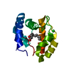
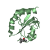
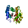
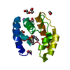
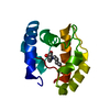
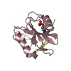
THE INHIBITOR (2,3-DIDEOXY-N,N',N'',N'''-TETRAACETYLCHITOTETRAOSE-1,5-LACTONE) IS COMPOSED OF SUGAR ...THE INHIBITOR (2,3-DIDEOXY-N,N',N'',N'''-TETRAACETYLCHITOTETRAOSE-1,5-LACTONE) IS COMPOSED OF SUGAR RESIDUES NAG 123, NAG 124, NAG 125, AND 4NN 126.
Sequence details
RESIDUE I5T IS NATURAL VARIANT ACCORDING TO REFERENCE 1 OF DATABASE UNIPROTKB/SWISS-PROT P86383 (LYS_MERLU).
-
Experimental details
-
Experiment
Experiment
Method: X-RAY DIFFRACTION / Number of used crystals: 1
-
Sample preparation
Crystal
Density Matthews: 2.04 Å3/Da / Density % sol: 39.58 %
Crystal grow
Temperature: 293 K / Method: vapor diffusion, sitting drop / pH: 5.5 Details: 35% 2-propanol, 0.1M Citrate buffer (pH5.5), 5% PEG 1000, VAPOR DIFFUSION, SITTING DROP, temperature 293K
In the structure databanks used in Yorodumi, some data are registered as the other names, "COVID-19 virus" and "2019-nCoV". Here are the details of the virus and the list of structure data.
Jan 31, 2019. EMDB accession codes are about to change! (news from PDBe EMDB page)
EMDB accession codes are about to change! (news from PDBe EMDB page)
The allocation of 4 digits for EMDB accession codes will soon come to an end. Whilst these codes will remain in use, new EMDB accession codes will include an additional digit and will expand incrementally as the available range of codes is exhausted. The current 4-digit format prefixed with “EMD-” (i.e. EMD-XXXX) will advance to a 5-digit format (i.e. EMD-XXXXX), and so on. It is currently estimated that the 4-digit codes will be depleted around Spring 2019, at which point the 5-digit format will come into force.
The EM Navigator/Yorodumi systems omit the EMD- prefix.
Related info.:Q: What is EMD? / ID/Accession-code notation in Yorodumi/EM Navigator
Yorodumi is a browser for structure data from EMDB, PDB, SASBDB, etc.
This page is also the successor to EM Navigator detail page, and also detail information page/front-end page for Omokage search.
The word "yorodu" (or yorozu) is an old Japanese word meaning "ten thousand". "mi" (miru) is to see.
Related info.:EMDB / PDB / SASBDB / Comparison of 3 databanks / Yorodumi Search / Aug 31, 2016. New EM Navigator & Yorodumi / Yorodumi Papers / Jmol/JSmol / Function and homology information / Changes in new EM Navigator and Yorodumi
 Movie
Movie Controller

 Yorodumi
 Open data
 Basic information
 Components
 Keywords
 Function and homology information
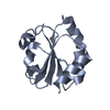
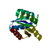
 Meretrix lusoria (invertebrata)
 X-RAY DIFFRACTION /
  Authors
 Citation
 Structure visualization
 Downloads & links
 Other downloads
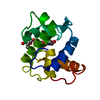

 PDBj
PDBj Assembly
 Mass: 18.015 Da / Num. of mol.: 148 / Source method: isolated from a natural source / Formula: H2O
 Sample preparation
 / Beamline: AR-NE3A / Wavelength: 1 Å
 Processing