 Movie
Movie Controller

[English] 日本語
 Yorodumi
- PDB-3fce: Crystal Structure of Bacillus cereus D-alanyl Carrier Protein Lig... -
+ Open data

- Basic information
| Entry | Database: PDB / ID: 3fce | ||||||
|---|---|---|---|---|---|---|---|
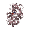
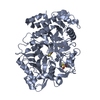
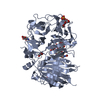
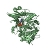
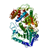
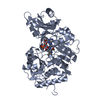
| Title | Crystal Structure of Bacillus cereus D-alanyl Carrier Protein Ligase DltA in Complex with ATP: Implications for Adenylation Mechanism | ||||||
 Components Components | D-alanine--poly(phosphoribitol) ligase subunit 1 | ||||||
 Keywords Keywords | LIGASE / DLTA / AMP-FORMING DOMAIN / D-ALANINE / ADENYLATION / D-ALANINE CARRIER PROTEIN LIGASE / ATP Complex | ||||||
| Function / homology |  Function and homology information Function and homology informationD-alanine-[D-alanyl-carrier protein] ligase / D-alanine [D-alanyl carrier protein] ligase activity / lipoteichoic acid biosynthetic process / ATP binding / cytoplasm Similarity search - Function | ||||||
| Biological species |  | ||||||
| Method |  X-RAY DIFFRACTION / FOURIER SYNTHESIS / Resolution: 1.9 Å X-RAY DIFFRACTION / FOURIER SYNTHESIS / Resolution: 1.9 Å | ||||||
 Authors Authors | Osman, K.T. / Du, L. / He, Y. / Luo, Y. | ||||||
 Citation Citation | Journal: J.Mol.Biol. / Year: 2009 Title: Crystal structure of Bacillus cereus D-alanyl carrier protein ligase (DltA) in complex with ATP. Authors: Osman, K.T. / Du, L. / He, Y. / Luo, Y. | ||||||
| History |
|
- Structure visualization
| Structure viewer | Molecule: MolmilJmol/JSmol |
|---|
- Downloads & links
-Download
| PDBx/mmCIF format | 3fce.cif.gz | 123.2 KB | Display | PDBx/mmCIF format |
|---|---|---|---|---|
| PDB format | pdb3fce.ent.gz | 92.1 KB | Display | PDB format |
| PDBx/mmJSON format | 3fce.json.gz | Tree view | PDBx/mmJSON format | |
| Others |  Other downloads Other downloads |
-Validation report
| Arichive directory | https://data.pdbj.org/pub/pdb/validation_reports/fc/3fceftp://data.pdbj.org/pub/pdb/validation_reports/fc/3fce | HTTPS FTP |
|---|
-Related structure data
| Related structure data | 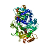 3fccC 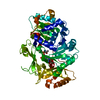 3dhvS S: Starting model for refinement C: citing same article ( |
|---|---|
| Similar structure data |


-Links
 PDBj
PDBj
- Assembly
| Deposited unit | 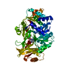
| ||||||||
|---|---|---|---|---|---|---|---|---|---|
| 1 |
| ||||||||
| Unit cell |
|
-Components
| #1: Protein | Mass: 57625.719 Da / Num. of mol.: 1 Source method: isolated from a genetically manipulated source Details: DltA was expressed as a fusion protein with C-terminal Histag. Source: (gene. exp.) |
|---|---|
| #2: Chemical | ChemComp-CA /   Mass: 40.078 Da / Num. of mol.: 1 / Source method: obtained synthetically / Formula: Ca Mass: 40.078 Da / Num. of mol.: 1 / Source method: obtained synthetically / Formula: Ca |
| #3: Chemical | ChemComp-ATP /   Mass: 507.181 Da / Num. of mol.: 1 / Source method: obtained synthetically / Formula: C10H16N5O13P3 / Comment: ATP, energy-carrying molecule*YM Mass: 507.181 Da / Num. of mol.: 1 / Source method: obtained synthetically / Formula: C10H16N5O13P3 / Comment: ATP, energy-carrying molecule*YM |
| #4: Water | ChemComp-HOH /  Mass: 18.015 Da / Num. of mol.: 424 / Source method: isolated from a natural source / Formula: H2O Mass: 18.015 Da / Num. of mol.: 424 / Source method: isolated from a natural source / Formula: H2O |
-Experimental details
-Experiment
| Experiment | Method: X-RAY DIFFRACTION / Number of used crystals: 1 |
|---|
- Sample preparation
| Crystal | Density Matthews: 2.41 Å3/Da / Density % sol: 48.88 % |
|---|---|
| Crystal grow | Temperature: 294 K / Method: vapor diffusion, hanging drop / pH: 7.2 Details: 0.05 M HEPES, 0.2 M POTASSIUM CHLORIDE, 0.1 M CALCIUM CHLORIDE, 16% PEG 3350, 21% sucrose, PH 7.2, VAPOR DIFFUSION, HANGING DROP, TEMPERATURE 294K |
-Data collection
| Diffraction | Mean temperature: 100 K |
|---|---|
| Diffraction source | Source: ROTATING ANODE / Type: BRUKER AXS MICROSTAR / Wavelength: 1.5418 / Wavelength: 1.5418 Å |
| Detector | Type: BRUKER SMART 6000 / Detector: CCD / Date: Oct 25, 2008 / Details: MIRRORS |
| Radiation | Monochromator: GRAPHITE / Protocol: SINGLE WAVELENGTH / Monochromatic (M) / Laue (L): M / Scattering type: x-ray |
| Radiation wavelength | Wavelength: 1.5418 Å / Relative weight: 1 |
| Reflection | Resolution: 1.9→20 Å / Num. all: 44062 / Num. obs: 38547 / % possible obs: 87.6 % / Observed criterion σ(F): 0 / Observed criterion σ(I): 0 / Redundancy: 6.4 % / Rmerge(I) obs: 0.06 / Rsym value: 0.06 |
| Reflection shell | Resolution: 1.9→2.02 Å / Redundancy: 5.1 % / Rmerge(I) obs: 0.306 / Mean I/σ(I) obs: 2 / Rsym value: 0.306 / % possible all: 44.7 |
- Processing
| Software |
| |||||||||||||||||||||||||
|---|---|---|---|---|---|---|---|---|---|---|---|---|---|---|---|---|---|---|---|---|---|---|---|---|---|---|
| Refinement | Method to determine structure: FOURIER SYNTHESIS Starting model: PDB ENTRY 3DHV Resolution: 1.9→20 Å / Cross valid method: THROUGHOUT / σ(F): 0 / Stereochemistry target values: Engh & Huber
| |||||||||||||||||||||||||
| Refinement step | Cycle: LAST / Resolution: 1.9→20 Å
| |||||||||||||||||||||||||
| Refine LS restraints |
|
