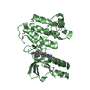
- PDB-3f82: Crystal structure of the tyrosine kinase domain of the hepatocyte... -
+
Open data
ID or keywords:
Loading...
-
Basic information
Entry
Database: PDB / ID: 3f82
Title
Crystal structure of the tyrosine kinase domain of the hepatocyte growth factor receptor C-MET in complex with N-(4-(2-amino-3-chloropyridin-4-yloxy)-3-fluorophenyl)-4-ethoxy-1-(4-fluorophenyl)-2-oxo-1,2-dihydropyridine-3-carboxamide
Protocol: SINGLE WAVELENGTH / Monochromatic (M) / Laue (L): M / Scattering type: x-ray
Radiation wavelength
Wavelength: 1 Å / Relative weight: 1
Reflection
Resolution: 2.5→25 Å / Num. obs: 11610 / % possible obs: 99.4 % / Observed criterion σ(I): 0 / Redundancy: 4.3 % / Biso Wilson estimate: 39.628 Å2 / Rmerge(I) obs: 0.079 / Net I/σ(I): 17.3
Reflection shell
Resolution: 2.5→2.59 Å / Redundancy: 3.7 % / Rmerge(I) obs: 0.275 / Mean I/σ(I) obs: 4.2 / % possible all: 97.2
-
Processing
Software
Name
Version
Classification
AMoRE
phasing
BUSTER-TNT
2.5.1
refinement
HKL-2000
(DENZO)
datareduction
HKL-2000
(SCALEPACK)
datascaling
Refinement
Method to determine structure: MOLECULAR REPLACEMENT Starting model: MET KINASE COMPLEXED WITH BMS-758982 Resolution: 2.5→16.54 Å / Cross valid method: THROUGHOUT / σ(F): 0
Rfactor
Num. reflection
% reflection
Selection details
Rfree
0.2533
547
4.79 %
RANDOM
Rwork
0.18
-
-
-
all
0.1834
11431
-
-
obs
0.1834
11431
99.31 %
-
Displacement parameters
Biso mean: 30.91 Å2
Baniso -1
Baniso -2
Baniso -3
1-
3.44456646 Å2
0 Å2
0 Å2
2-
-
-0.2538769 Å2
0 Å2
3-
-
-
-3.19068956 Å2
Refinement step
Cycle: LAST / Resolution: 2.5→16.54 Å
Protein
Nucleic acid
Ligand
Solvent
Total
Num. atoms
2169
0
36
127
2332
Refine LS restraints
Refine-ID
Type
Dev ideal
Number
Weight
X-RAY DIFFRACTION
t_bond_d
0.006
2261
2
X-RAY DIFFRACTION
t_angle_deg
0.868
3054
2
X-RAY DIFFRACTION
t_dihedral_angle_d
21.361
422
0
X-RAY DIFFRACTION
t_incorr_chiral_ct
X-RAY DIFFRACTION
t_pseud_angle
X-RAY DIFFRACTION
t_trig_c_planes
0.006
40
2
X-RAY DIFFRACTION
t_gen_planes
0.01
326
5
X-RAY DIFFRACTION
t_it
1.319
2261
20
X-RAY DIFFRACTION
t_nbd
0.032
29
5
LS refinement shell
Resolution: 2.5→2.65 Å / Total num. of bins used: 9
Rfactor
Num. reflection
% reflection
Rfree
0.2578
80
4.54 %
Rwork
0.1825
1682
-
all
0.186
1762
-
obs
-
-
99.31 %
+
About Yorodumi
-
News
-
Feb 9, 2022. New format data for meta-information of EMDB entries
New format data for meta-information of EMDB entries
Version 3 of the EMDB header file is now the official format.
The previous official version 1.9 will be removed from the archive.
In the structure databanks used in Yorodumi, some data are registered as the other names, "COVID-19 virus" and "2019-nCoV". Here are the details of the virus and the list of structure data.
Jan 31, 2019. EMDB accession codes are about to change! (news from PDBe EMDB page)
EMDB accession codes are about to change! (news from PDBe EMDB page)
The allocation of 4 digits for EMDB accession codes will soon come to an end. Whilst these codes will remain in use, new EMDB accession codes will include an additional digit and will expand incrementally as the available range of codes is exhausted. The current 4-digit format prefixed with “EMD-” (i.e. EMD-XXXX) will advance to a 5-digit format (i.e. EMD-XXXXX), and so on. It is currently estimated that the 4-digit codes will be depleted around Spring 2019, at which point the 5-digit format will come into force.
The EM Navigator/Yorodumi systems omit the EMD- prefix.
Related info.:Q: What is EMD? / ID/Accession-code notation in Yorodumi/EM Navigator
Yorodumi is a browser for structure data from EMDB, PDB, SASBDB, etc.
This page is also the successor to EM Navigator detail page, and also detail information page/front-end page for Omokage search.
The word "yorodu" (or yorozu) is an old Japanese word meaning "ten thousand". "mi" (miru) is to see.
Related info.:EMDB / PDB / SASBDB / Comparison of 3 databanks / Yorodumi Search / Aug 31, 2016. New EM Navigator & Yorodumi / Yorodumi Papers / Jmol/JSmol / Function and homology information / Changes in new EM Navigator and Yorodumi
 Movie
Movie Controller

 Yorodumi
 Open data
 Basic information
 Components
 Keywords
 Function and homology information
 Homo sapiens (human)
 X-RAY DIFFRACTION /
  Authors
 Citation
 Structure visualization
 Downloads & links
 Other downloads
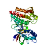


 PDBj
PDBj


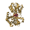
 Assembly

 Spodoptera frugiperda (fall armyworm)
 Mass: 512.893 Da / Num. of mol.: 1 / Source method: obtained synthetically / Formula: C25H19ClF2N4O4
 Mass: 18.015 Da / Num. of mol.: 127 / Source method: isolated from a natural source / Formula: H2O
 Sample preparation
 / Beamline: 17-ID / Wavelength: 1
  Processing