Mass: 18.015 Da / Num. of mol.: 462 / Source method: isolated from a natural source / Formula: H2O
Nonpolymer details
1. 8PA IS A COVALENT ADDUCT BETWEEN 3-(PYRIDIN-3-YL) ACRYLALDEHYDE AND THE THDP COFACTOR. 2. ...1. 8PA IS A COVALENT ADDUCT BETWEEN 3-(PYRIDIN-3-YL) ACRYLALDEHYDE AND THE THDP COFACTOR. 2. ACCORDING TO AUTHORS, BOTH RELATED ENTRIES 3F6B AND 3F6E HAVE THE SAME SUBSTRATE-COFACTOR ADDUCT. THESE STRUCTURES WERE SOLVED IN SUPPORT OF ENZYMOLOGICAL EXPERIMENTS WITH BENZOYLFORMATE DECARBOXYLASE. THE ENZYME DECARBOXYLATES THE 3-PKB SUBSTRATE TO GENERATE AN ALDEHYDE. THIS ALDEHYDE IS PAA, THE SUBSTRATE APPLIED IN THE EXPERIMENTS THAT GAVE RISE TO 3F6B STRUCTURE. WHEN AUTHORS CRYSTALLIZE THE ENZYME IN THE PRESENCE OF 3-PKB, THE SPECIES THAT IS GENERATED THAN TRAPPED IS PAA, VIA REACTION WITH THE THIAZOLIUM YLID OF THE THDP COFACTOR TO GENERATE THE ADDUCT SEEN IN BOTH STRUCTURES. THE 3-PKB INHIBITOR WAS DESIGNED FIRST, AND THEN PAA WAS DEVELOPED TO CHECK THE HYPOTHESIS THAT 3-PKB IS CONVERTED TO PAA IN THE ACTIVE SITE. INDEED, THE ADDUCT IS THE SAME IN 3F6B AND 3F6E.
-
Experimental details
-
Experiment
Experiment
Method: X-RAY DIFFRACTION / Number of used crystals: 1
-
Sample preparation
Crystal
Density Matthews: 2.4 Å3/Da / Density % sol: 48.75 %
Crystal grow
Temperature: 298 K / Method: vapor diffusion, hanging drop / pH: 8.5 Details: 100 mM Tris-HCl pH 8.5, 150 mM CaCl2, 0.5% v/v MPD (2-methyl-2,4-pentanediol), 22% v/v PEG 400, VAPOR DIFFUSION, HANGING DROP, temperature 298K
In the structure databanks used in Yorodumi, some data are registered as the other names, "COVID-19 virus" and "2019-nCoV". Here are the details of the virus and the list of structure data.
Jan 31, 2019. EMDB accession codes are about to change! (news from PDBe EMDB page)
EMDB accession codes are about to change! (news from PDBe EMDB page)
The allocation of 4 digits for EMDB accession codes will soon come to an end. Whilst these codes will remain in use, new EMDB accession codes will include an additional digit and will expand incrementally as the available range of codes is exhausted. The current 4-digit format prefixed with “EMD-” (i.e. EMD-XXXX) will advance to a 5-digit format (i.e. EMD-XXXXX), and so on. It is currently estimated that the 4-digit codes will be depleted around Spring 2019, at which point the 5-digit format will come into force.
The EM Navigator/Yorodumi systems omit the EMD- prefix.
Related info.:Q: What is EMD? / ID/Accession-code notation in Yorodumi/EM Navigator
Yorodumi is a browser for structure data from EMDB, PDB, SASBDB, etc.
This page is also the successor to EM Navigator detail page, and also detail information page/front-end page for Omokage search.
The word "yorodu" (or yorozu) is an old Japanese word meaning "ten thousand". "mi" (miru) is to see.
Related info.:EMDB / PDB / SASBDB / Comparison of 3 databanks / Yorodumi Search / Aug 31, 2016. New EM Navigator & Yorodumi / Yorodumi Papers / Jmol/JSmol / Function and homology information / Changes in new EM Navigator and Yorodumi
 Movie
Movie Controller

 Yorodumi
 Open data
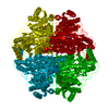
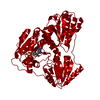
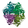
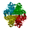
 Basic information
 Components
 Keywords
 Function and homology information
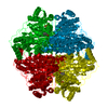
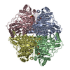
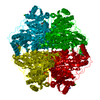
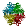
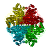
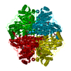
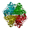
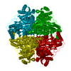
 Pseudomonas putida (bacteria)
 X-RAY DIFFRACTION /
  Authors
 Citation
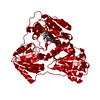
 Structure visualization
 Downloads & links
 Other downloads


 PDBj
PDBj


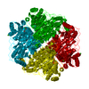
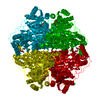
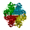
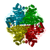
 Assembly
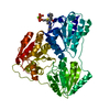


 Mass: 24.305 Da / Num. of mol.: 1 / Source method: obtained synthetically / Formula: Mg
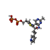
 Mass: 558.462 Da / Num. of mol.: 1 / Source method: obtained synthetically / Formula: C20H26N5O8P2S
 Mass: 18.015 Da / Num. of mol.: 462 / Source method: isolated from a natural source / Formula: H2O
 Sample preparation
 / Beamline: 23-ID-D / Wavelength: 1.0332 Å
 Processing