 Movie
Movie Controller

[English] 日本語
 Yorodumi
- PDB-1q6z: HIGH RESOLUTION STRUCTURE OF E28A MUTANT BENZOYLFORMATE DECARBOXY... -
+ Open data

- Basic information
| Entry | Database: PDB / ID: 1q6z | ||||||
|---|---|---|---|---|---|---|---|
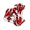
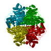
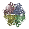
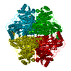
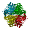
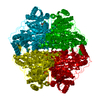
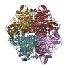
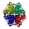
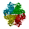
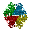
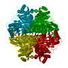
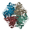
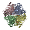
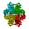
| Title | HIGH RESOLUTION STRUCTURE OF E28A MUTANT BENZOYLFORMATE DECARBOXYLASE FROM PSEUDOMONAS PUTIDA COMPLEXED WITH THIAMIN THIAZOLONE DIPHOSPHATE | ||||||
 Components Components | Benzoylformate decarboxylase | ||||||
 Keywords Keywords | LYASE / CARBON-CARBON / DECARBOXYLASE / MANDELATE CATABOLISM / THIAMIN THIAZOLONE DIPHOSPHATE / INHIBITOR / HIGH RESOLUTION | ||||||
| Function / homology |  Function and homology information Function and homology informationbenzoylformate decarboxylase / benzoylformate decarboxylase activity / (R)-mandelate catabolic process / acetolactate synthase activity / thiamine pyrophosphate binding / flavin adenine dinucleotide binding / magnesium ion binding Similarity search - Function | ||||||
| Biological species |  Pseudomonas putida (bacteria) Pseudomonas putida (bacteria) | ||||||
| Method |  X-RAY DIFFRACTION / SYNCHROTRON / MOLECULAR REPLACEMENT / Resolution: 1 Å X-RAY DIFFRACTION / SYNCHROTRON / MOLECULAR REPLACEMENT / Resolution: 1 Å | ||||||
 Authors Authors | Bera, A.K. / Hasson, M.S. | ||||||
 Citation Citation | Journal: To be Published Title: High Resolution Structure of E28A Mutant Benzoylformate Decarboxylase from Pseudomonas Putida Complexed with Thiamin Thiazolone Diphosphate Authors: Bera, A.K. / Anderson, N.L. / Hasson, M.S. #1: Journal: Biochemistry / Year: 1998Title: The Crystal Structure of Benzoylformate Decarboxylase at 1.6 A Resolution: Diversity of Catalytic Residues in Thiamin Diphosphate-Dependent Enzymes Authors: Hasson, M.S. / Muscate, A. / Mcleish, M.J. / Polovnikova, L.S. / Gerlt, J.A. / Kenyon, G.L. / Petsko, G.A. / Ringe, D. #2: Journal: Biochemistry / Year: 2003Title: Structural and Kinetic Analysis of Catalysis by a Thiamin Diphosphate-Dependent Enzyme, Benzoylformate Decarboxylase Authors: Polovnikova, E.S. / Mcleish, M.J. / Sergienko, E.A. / Burgner, J.T. / Anderson, N.L. / Bera, A.K. / Jordan, F. / Kenyon, G.L. / Hasson, M.S. | ||||||
| History |
|
- Structure visualization
| Structure viewer | Molecule: MolmilJmol/JSmol |
|---|
- Downloads & links
-Download
| PDBx/mmCIF format | 1q6z.cif.gz | 131.1 KB | Display | PDBx/mmCIF format |
|---|---|---|---|---|
| PDB format | pdb1q6z.ent.gz | 97.7 KB | Display | PDB format |
| PDBx/mmJSON format | 1q6z.json.gz | Tree view | PDBx/mmJSON format | |
| Others |  Other downloads Other downloads |
-Validation report
| Arichive directory | https://data.pdbj.org/pub/pdb/validation_reports/q6/1q6zftp://data.pdbj.org/pub/pdb/validation_reports/q6/1q6z | HTTPS FTP |
|---|
-Related structure data
| Related structure data |  1bfdS S: Starting model for refinement |
|---|---|
| Similar structure data |


-Links
 PDBj
PDBj


- Assembly
| Deposited unit | 
| ||||||||||||||||||
|---|---|---|---|---|---|---|---|---|---|---|---|---|---|---|---|---|---|---|---|
| 1 | 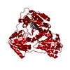
| ||||||||||||||||||
| Unit cell |
| ||||||||||||||||||
| Components on special symmetry positions |
|
-Components
| #1: Protein | Mass: 56344.715 Da / Num. of mol.: 1 / Mutation: E28A Source method: isolated from a genetically manipulated source Source: (gene. exp.) Pseudomonas putida (bacteria) / Strain: RF4738 / Gene: MDLC / Plasmid: pRep7 / Gene (production host): MDLC / Production host: | ||||
|---|---|---|---|---|---|
| #2: Chemical | ChemComp-MG /   Mass: 24.305 Da / Num. of mol.: 1 / Source method: obtained synthetically / Formula: Mg Mass: 24.305 Da / Num. of mol.: 1 / Source method: obtained synthetically / Formula: Mg | ||||
| #3: Chemical |   Mass: 40.078 Da / Num. of mol.: 3 / Source method: obtained synthetically / Formula: Ca Mass: 40.078 Da / Num. of mol.: 3 / Source method: obtained synthetically / Formula: Ca#4: Chemical | ChemComp-TZD / |   Mass: 440.306 Da / Num. of mol.: 1 / Source method: obtained synthetically / Formula: C12H18N4O8P2S Mass: 440.306 Da / Num. of mol.: 1 / Source method: obtained synthetically / Formula: C12H18N4O8P2S#5: Water | ChemComp-HOH / |  Mass: 18.015 Da / Num. of mol.: 588 / Source method: isolated from a natural source / Formula: H2O Mass: 18.015 Da / Num. of mol.: 588 / Source method: isolated from a natural source / Formula: H2O |
-Experimental details
-Experiment
| Experiment | Method: X-RAY DIFFRACTION / Number of used crystals: 1 |
|---|
- Sample preparation
| Crystal | Density Matthews: 2.04 Å3/Da / Density % sol: 47.89 % |
|---|---|
| Crystal grow | Temperature: 293 K / Method: vapor diffusion, hanging drop / pH: 8.5 Details: pH 8.5, VAPOR DIFFUSION, HANGING DROP, temperature 293K |
-Data collection
| Diffraction | Mean temperature: 100 K | |||||||||
|---|---|---|---|---|---|---|---|---|---|---|
| Diffraction source | Source: SYNCHROTRON / Site: APS  / Beamline: 14-BM-C / Wavelength: 0.9 / Wavelength: 0.9 Å / Beamline: 14-BM-C / Wavelength: 0.9 / Wavelength: 0.9 Å | |||||||||
| Detector | Type: ADSC QUANTUM 4 / Detector: CCD / Date: Jul 1, 2003 | |||||||||
| Radiation | Protocol: SINGLE WAVELENGTH / Monochromatic (M) / Laue (L): M / Scattering type: x-ray | |||||||||
| Radiation wavelength |
| |||||||||
| Reflection | Resolution: 1→30 Å / Num. obs: 242762 / % possible obs: 89.3 % / Observed criterion σ(I): 0 / Rmerge(I) obs: 0.053 / Net I/σ(I): 19 | |||||||||
| Reflection shell | Resolution: 1→1.03 Å / Rmerge(I) obs: 0.336 / % possible all: 40.7 |
- Processing
| Software |
| |||||||||||||||||||||||||||||||||
|---|---|---|---|---|---|---|---|---|---|---|---|---|---|---|---|---|---|---|---|---|---|---|---|---|---|---|---|---|---|---|---|---|---|---|
| Refinement | Method to determine structure: MOLECULAR REPLACEMENT Starting model: PDB ENTRY 1BFD Resolution: 1→30 Å / Num. parameters: 41647 / Num. restraintsaints: 49689 / Cross valid method: FREE R / σ(F): 0 / Stereochemistry target values: ENGH AND HUBER
| |||||||||||||||||||||||||||||||||
| Refine analyze | Num. disordered residues: 9 / Occupancy sum hydrogen: 3771.56 / Occupancy sum non hydrogen: 4419.85 | |||||||||||||||||||||||||||||||||
| Refinement step | Cycle: LAST / Resolution: 1→30 Å
| |||||||||||||||||||||||||||||||||
| Refine LS restraints |
|
