 Movie
Movie Controller


+ Open data

- Basic information
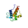
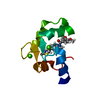
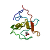
| Entry | Database: PDB / ID: 3f2e | ||||||
|---|---|---|---|---|---|---|---|
| Title | Crystal structure of Yellowstone SIRV coat protein C-terminus | ||||||
 Components Components | SIRV coat protein | ||||||
 Keywords Keywords | VIRAL PROTEIN / four helix bundle / virus coat protein | ||||||
| Function / homology | Sulfolobus virus, coat protein, C-terminal / Sulfolobus virus coat protein C terminal / Methane Monooxygenase Hydroxylase; Chain G, domain 1 - #800 / Methane Monooxygenase Hydroxylase; Chain G, domain 1 / Up-down Bundle / Mainly Alpha / CITRIC ACID / SIRV coat protein Function and homology information Function and homology information | ||||||
| Biological species |   Sulfolobus islandicus rudivirus 1 variant YNP Sulfolobus islandicus rudivirus 1 variant YNP | ||||||
| Method |  X-RAY DIFFRACTION / MOLECULAR REPLACEMENT / Resolution: 1.668 Å X-RAY DIFFRACTION / MOLECULAR REPLACEMENT / Resolution: 1.668 Å | ||||||
 Authors Authors | Taurog, R.E. / Szymczyna, B.R. / Williamson, J.R. / Johnson, J.E. | ||||||
 Citation Citation | Journal: Structure / Year: 2009 Title: Synergy of NMR, computation, and X-ray crystallography for structural biology. Authors: Szymczyna, B.R. / Taurog, R.E. / Young, M.J. / Snyder, J.C. / Johnson, J.E. / Williamson, J.R. | ||||||
| History |
|
- Structure visualization
| Structure viewer | Molecule: MolmilJmol/JSmol |
|---|
- Downloads & links
-Download
| PDBx/mmCIF format | 3f2e.cif.gz | 29.5 KB | Display | PDBx/mmCIF format |
|---|---|---|---|---|
| PDB format | pdb3f2e.ent.gz | 18.9 KB | Display | PDB format |
| PDBx/mmJSON format | 3f2e.json.gz | Tree view | PDBx/mmJSON format | |
| Others |  Other downloads Other downloads |
-Validation report
| Arichive directory | https://data.pdbj.org/pub/pdb/validation_reports/f2/3f2eftp://data.pdbj.org/pub/pdb/validation_reports/f2/3f2e | HTTPS FTP |
|---|
-Related structure data
| Similar structure data |
|---|


-Links
 PDBj
PDBj- Assembly
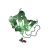
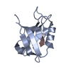
| Deposited unit | 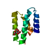
| ||||||||
|---|---|---|---|---|---|---|---|---|---|
| 1 |
| ||||||||
| Unit cell |
|
-Components
| #1: Protein | Mass: 10498.848 Da / Num. of mol.: 1 / Fragment: C-terminal domain Source method: isolated from a genetically manipulated source Details: pET31a tags were removed from the vector, a C-terminal hexahistidine tag was cloned in Source: (gene. exp.) Sulfolobus islandicus rudivirus 1 variant YNPPlasmid: pET31a / Production host:  |
|---|---|
| #2: Chemical | ChemComp-CIT /   Mass: 192.124 Da / Num. of mol.: 1 / Source method: obtained synthetically / Formula: C6H8O7 Mass: 192.124 Da / Num. of mol.: 1 / Source method: obtained synthetically / Formula: C6H8O7 |
| #3: Water | ChemComp-HOH /  Mass: 18.015 Da / Num. of mol.: 80 / Source method: isolated from a natural source / Formula: H2O Mass: 18.015 Da / Num. of mol.: 80 / Source method: isolated from a natural source / Formula: H2O |
-Experimental details
-Experiment
| Experiment | Method: X-RAY DIFFRACTION / Number of used crystals: 1 |
|---|
- Sample preparation
| Crystal | Density Matthews: 2.74 Å3/Da / Density % sol: 55.14 % |
|---|---|
| Crystal grow | Temperature: 299 K / Method: vapor diffusion, sitting drop Details: SIRV CP (residues 46-134) at 19 or 30 mg/ml in 20 mM Na MES pH 6.0 was mixed 1:1 with 25% PEG 20,000, 0.1 M Na Citrate pH 3.6, 4-9% sucrose. Crystals were frozen in of 14% PEG 20,000, 50 mM ...Details: SIRV CP (residues 46-134) at 19 or 30 mg/ml in 20 mM Na MES pH 6.0 was mixed 1:1 with 25% PEG 20,000, 0.1 M Na Citrate pH 3.6, 4-9% sucrose. Crystals were frozen in of 14% PEG 20,000, 50 mM sodium citrate pH 3.6, 15-25% sucrose, VAPOR DIFFUSION, SITTING DROP, temperature 299K |
-Data collection
| Diffraction | Mean temperature: 173 K |
|---|---|
| Diffraction source | Source: ROTATING ANODE / Type: RIGAKU FR-D / Wavelength: 1.542 Å |
| Detector | Type: MAR scanner 345 mm plate / Detector: IMAGE PLATE / Date: Aug 12, 2008 |
| Radiation | Protocol: SINGLE WAVELENGTH / Monochromatic (M) / Laue (L): M / Scattering type: x-ray |
| Radiation wavelength | Wavelength: 1.542 Å / Relative weight: 1 |
| Reflection | Resolution: 1.668→44.588 Å / Num. obs: 14062 / % possible obs: 99.1 % / Observed criterion σ(I): -3 / Redundancy: 16.8 % / Biso Wilson estimate: 24.675 Å2 / Rmerge(I) obs: 0.034 / Rsym value: 0.062 / Net I/σ(I): 33.4 |
| Reflection shell | Resolution: 1.668→1.7 Å / Redundancy: 14.5 % / Rmerge(I) obs: 0.203 / Mean I/σ(I) obs: 8.27 / Num. unique all: 965 / Rsym value: 0.371 / % possible all: 94.6 |
- Processing
| Software |
| ||||||||||||||||||||||||||||||||||||||||||
|---|---|---|---|---|---|---|---|---|---|---|---|---|---|---|---|---|---|---|---|---|---|---|---|---|---|---|---|---|---|---|---|---|---|---|---|---|---|---|---|---|---|---|---|
| Refinement | Method to determine structure: MOLECULAR REPLACEMENT Starting model: CS-Rosetta model Resolution: 1.668→31.642 Å / SU ML: 0.14 / Isotropic thermal model: isotropic / Cross valid method: THROUGHOUT / σ(F): 2 / Stereochemistry target values: ML
| ||||||||||||||||||||||||||||||||||||||||||
| Solvent computation | Shrinkage radii: 0.9 Å / VDW probe radii: 1.11 Å / Solvent model: FLAT BULK SOLVENT MODEL / Bsol: 49.566 Å2 / ksol: 0.39 e/Å3 | ||||||||||||||||||||||||||||||||||||||||||
| Displacement parameters | Biso mean: 20.5 Å2
| ||||||||||||||||||||||||||||||||||||||||||
| Refine analyze | Luzzati coordinate error obs: 0.177 Å | ||||||||||||||||||||||||||||||||||||||||||
| Refinement step | Cycle: LAST / Resolution: 1.668→31.642 Å
| ||||||||||||||||||||||||||||||||||||||||||
| Refine LS restraints |
| ||||||||||||||||||||||||||||||||||||||||||
| LS refinement shell | Refine-ID: X-RAY DIFFRACTION
|
