cardiogenic plate morphogenesis / endodermal cell fate determination / regulation of cardiac cell fate specification / stem cell fate specification / endocardium formation / common bile duct development / ureter development / endodermal digestive tract morphogenesis / gallbladder development / inner cell mass cellular morphogenesis ...cardiogenic plate morphogenesis / endodermal cell fate determination / regulation of cardiac cell fate specification / stem cell fate specification / endocardium formation / common bile duct development / ureter development / endodermal digestive tract morphogenesis / gallbladder development / inner cell mass cellular morphogenesis / cardiac cell fate determination / endodermal cell fate specification / regulation of stem cell division / cell migration involved in gastrulation / Deactivation of the beta-catenin transactivating complex / endocardial cell differentiation / rostrocaudal neural tube patterning / positive regulation of endodermal cell differentiation / positive regulation of stem cell differentiation / embryonic foregut morphogenesis / regulation of stem cell proliferation / endoderm development / response to alkaloid / metanephros development / embryonic heart tube morphogenesis / embryonic heart tube development / signal transduction involved in regulation of gene expression / heart looping / negative regulation of Wnt signaling pathway / endodermal cell differentiation / regulation of cell differentiation / outflow tract morphogenesis / regulation of embryonic development / embryonic organ development / vasculogenesis / gastrulation / cellular response to leukemia inhibitory factor / stem cell differentiation / negative regulation of canonical Wnt signaling pathway / negative regulation of cell growth / protein destabilization / beta-catenin binding / Wnt signaling pathway / positive regulation of protein catabolic process / heart development / DNA-binding transcription activator activity, RNA polymerase II-specific / angiogenesis / spermatogenesis / transcription regulator complex / gene expression / sequence-specific DNA binding / RNA polymerase II-specific DNA-binding transcription factor binding / DNA-binding transcription factor activity, RNA polymerase II-specific / transcription cis-regulatory region binding / protein stabilization / DNA-binding transcription factor activity / positive regulation of gene expression / regulation of DNA-templated transcription / regulation of transcription by RNA polymerase II / positive regulation of DNA-templated transcription / positive regulation of transcription by RNA polymerase II / DNA binding / nucleoplasm / nucleus Similarity search - Function
Sox, C-terminal / Sox 7/17/18, central domain / Sox 17/18 central domain / Sox C-terminal domain profile. / : / High mobility group box domain / DNA Binding (I), subunit A / HMG (high mobility group) box / HMG boxes A and B DNA-binding domains profile. / high mobility group ...Sox, C-terminal / Sox 7/17/18, central domain / Sox 17/18 central domain / Sox C-terminal domain profile. / : / High mobility group box domain / DNA Binding (I), subunit A / HMG (high mobility group) box / HMG boxes A and B DNA-binding domains profile. / high mobility group / High mobility group box domain / High mobility group box domain superfamily / Orthogonal Bundle / Mainly Alpha Similarity search - Domain/homology
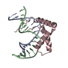
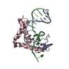
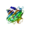
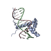
A: DNA (5'-D(*DGP*DTP*DCP*DTP*DCP*DTP*DAP*DTP*DTP*DGP*DTP*DCP*DCP*DTP*DGP*DG)-3') B: DNA (5'-D(*DCP*DCP*DAP*DGP*DGP*DAP*DCP*DAP*DAP*DTP*DAP*DGP*DAP*DGP*DAP*DC)-3') D: Transcription factor SOX-17
In the structure databanks used in Yorodumi, some data are registered as the other names, "COVID-19 virus" and "2019-nCoV". Here are the details of the virus and the list of structure data.
Jan 31, 2019. EMDB accession codes are about to change! (news from PDBe EMDB page)
EMDB accession codes are about to change! (news from PDBe EMDB page)
The allocation of 4 digits for EMDB accession codes will soon come to an end. Whilst these codes will remain in use, new EMDB accession codes will include an additional digit and will expand incrementally as the available range of codes is exhausted. The current 4-digit format prefixed with “EMD-” (i.e. EMD-XXXX) will advance to a 5-digit format (i.e. EMD-XXXXX), and so on. It is currently estimated that the 4-digit codes will be depleted around Spring 2019, at which point the 5-digit format will come into force.
The EM Navigator/Yorodumi systems omit the EMD- prefix.
Related info.:Q: What is EMD? / ID/Accession-code notation in Yorodumi/EM Navigator
Yorodumi is a browser for structure data from EMDB, PDB, SASBDB, etc.
This page is also the successor to EM Navigator detail page, and also detail information page/front-end page for Omokage search.
The word "yorodu" (or yorozu) is an old Japanese word meaning "ten thousand". "mi" (miru) is to see.
Related info.:EMDB / PDB / SASBDB / Comparison of 3 databanks / Yorodumi Search / Aug 31, 2016. New EM Navigator & Yorodumi / Yorodumi Papers / Jmol/JSmol / Function and homology information / Changes in new EM Navigator and Yorodumi
 Movie
Movie Controller


 Open data
 Basic information
 Components
 Keywords
 Function and homology information
 X-RAY DIFFRACTION /
  Authors
 Citation
 Structure visualization
 Downloads & links
 Other downloads
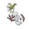


 PDBj
PDBj


 Assembly

 Sample preparation
 Processing