 Movie
Movie Controller

[English] 日本語
 Yorodumi
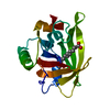
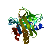
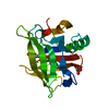
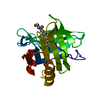
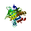
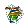
- PDB-3ew7: Crystal structure of the Lmo0794 protein from Listeria monocytoge... -
+ Open data

- Basic information
| Entry | Database: PDB / ID: 3ew7 | ||||||
|---|---|---|---|---|---|---|---|
| Title | Crystal structure of the Lmo0794 protein from Listeria monocytogenes. Northeast Structural Genomics Consortium target LmR162. | ||||||
 Components Components | Lmo0794 protein | ||||||
 Keywords Keywords | structural genomics / unknown function / Q8Y8U8_LISMO / Lmo0794 / putative NAD-dependent Epimerase/dehydratase / LmR162 / NESG / PSI-2 / Protein Structure Initiative / Northeast Structural Genomics Consortium | ||||||
| Function / homology |  Function and homology information Function and homology informationoxidoreductase activity, acting on the CH-NH group of donors, NAD or NADP as acceptor Similarity search - Function | ||||||
| Biological species |  Listeria monocytogenes (bacteria) Listeria monocytogenes (bacteria) | ||||||
| Method |  X-RAY DIFFRACTION / SYNCHROTRON / MAD / Resolution: 2.73 Å X-RAY DIFFRACTION / SYNCHROTRON / MAD / Resolution: 2.73 Å | ||||||
 Authors Authors | Vorobiev, S.M. / Abashidze, M. / Seetharaman, J. / Wang, D. / Foote, E.L. / Ciccosanti, C. / Janjua, H. / Xiao, R. / Acton, T.B. / Montelione, G.T. ...Vorobiev, S.M. / Abashidze, M. / Seetharaman, J. / Wang, D. / Foote, E.L. / Ciccosanti, C. / Janjua, H. / Xiao, R. / Acton, T.B. / Montelione, G.T. / Tong, L. / Hunt, J.F. / Northeast Structural Genomics Consortium (NESG) | ||||||
 Citation Citation | Journal: To be Published Title: Crystal structure of the Lmo0794 protein from Listeria monocytogenes. Authors: Vorobiev, S.M. / Abashidze, M. / Seetharaman, J. / Wang, D. / Foote, E.L. / Ciccosanti, C. / Janjua, H. / Xiao, R. / Acton, T.B. / Montelione, G.T. / Tong, L. / Hunt, J.F. | ||||||
| History |
|
- Structure visualization
| Structure viewer | Molecule: MolmilJmol/JSmol |
|---|
- Downloads & links
-Download
| PDBx/mmCIF format | 3ew7.cif.gz | 47.6 KB | Display | PDBx/mmCIF format |
|---|---|---|---|---|
| PDB format | pdb3ew7.ent.gz | 33.1 KB | Display | PDB format |
| PDBx/mmJSON format | 3ew7.json.gz | Tree view | PDBx/mmJSON format | |
| Others |  Other downloads Other downloads |
-Validation report
| Arichive directory | https://data.pdbj.org/pub/pdb/validation_reports/ew/3ew7ftp://data.pdbj.org/pub/pdb/validation_reports/ew/3ew7 | HTTPS FTP |
|---|
-Related structure data
| Similar structure data | |
|---|---|
| Other databases |


-Links
 PDBj
PDBj- Assembly
| Deposited unit | 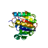
| ||||||||
|---|---|---|---|---|---|---|---|---|---|
| 1 |
| ||||||||
| Unit cell |
|
-Components
| #1: Protein | Mass: 24563.934 Da / Num. of mol.: 1 / Mutation: I71V, K142R, H207R Source method: isolated from a genetically manipulated source Source: (gene. exp.) Listeria monocytogenes (bacteria) / Strain: ATCC BAA-679/EGD-e/Serovar 1/2a / Gene: lmo0794 / Production host: |
|---|---|
| #2: Water | ChemComp-HOH /  Mass: 18.015 Da / Num. of mol.: 77 / Source method: isolated from a natural source / Formula: H2O Mass: 18.015 Da / Num. of mol.: 77 / Source method: isolated from a natural source / Formula: H2O |
| Has protein modification | Y |
-Experimental details
-Experiment
| Experiment | Method: X-RAY DIFFRACTION / Number of used crystals: 1 |
|---|
- Sample preparation
| Crystal | Density Matthews: 3.8 Å3/Da / Density % sol: 67.65 % |
|---|---|
| Crystal grow | Temperature: 292 K / Method: vapor diffusion, hanging drop / pH: 4.6 Details: 18% PEG 4000, 0.1M KH(2)PO(4), 0.1M sodium acetate, pH 4.6, VAPOR DIFFUSION, HANGING DROP, temperature 292K |
-Data collection
| Diffraction | Mean temperature: 100 K | ||||||||||||
|---|---|---|---|---|---|---|---|---|---|---|---|---|---|
| Diffraction source | Source: SYNCHROTRON / Site: NSLS  / Beamline: X4A / Wavelength: 0.97894, 0.97926, 0.96785 / Beamline: X4A / Wavelength: 0.97894, 0.97926, 0.96785 | ||||||||||||
| Detector | Type: ADSC QUANTUM 4 / Detector: CCD / Date: Aug 8, 2008 | ||||||||||||
| Radiation | Protocol: MAD / Monochromatic (M) / Laue (L): M / Scattering type: x-ray | ||||||||||||
| Radiation wavelength |
| ||||||||||||
| Reflection | Resolution: 2.73→50 Å / Num. all: 19317 / Num. obs: 19111 / % possible obs: 98.9 % / Observed criterion σ(F): 0 / Observed criterion σ(I): 0 / Redundancy: 4.5 % / Biso Wilson estimate: 47.1 Å2 / Rmerge(I) obs: 0.106 / Net I/σ(I): 16.05 | ||||||||||||
| Reflection shell | Resolution: 2.73→2.85 Å / Redundancy: 4.6 % / Rmerge(I) obs: 0.648 / Mean I/σ(I) obs: 4.05 / % possible all: 97.9 |
- Processing
| Software |
| ||||||||||||||||||||||||||||||||||||||||||||||||||||||||||||
|---|---|---|---|---|---|---|---|---|---|---|---|---|---|---|---|---|---|---|---|---|---|---|---|---|---|---|---|---|---|---|---|---|---|---|---|---|---|---|---|---|---|---|---|---|---|---|---|---|---|---|---|---|---|---|---|---|---|---|---|---|---|
| Refinement | Method to determine structure: MAD / Resolution: 2.73→33.76 Å / Rfactor Rfree error: 0.007 / Data cutoff high absF: 362304.23 / Data cutoff low absF: 0 / Isotropic thermal model: RESTRAINED / Cross valid method: THROUGHOUT / σ(F): 2 / Stereochemistry target values: Engh & Huber / Details: BULK SOLVENT MODEL USED
| ||||||||||||||||||||||||||||||||||||||||||||||||||||||||||||
| Solvent computation | Solvent model: FLAT MODEL / Bsol: 25.7195 Å2 / ksol: 0.3 e/Å3 | ||||||||||||||||||||||||||||||||||||||||||||||||||||||||||||
| Displacement parameters | Biso mean: 61.1 Å2
| ||||||||||||||||||||||||||||||||||||||||||||||||||||||||||||
| Refine analyze |
| ||||||||||||||||||||||||||||||||||||||||||||||||||||||||||||
| Refinement step | Cycle: LAST / Resolution: 2.73→33.76 Å
| ||||||||||||||||||||||||||||||||||||||||||||||||||||||||||||
| Refine LS restraints |
| ||||||||||||||||||||||||||||||||||||||||||||||||||||||||||||
| LS refinement shell | Resolution: 2.73→2.9 Å / Rfactor Rfree error: 0.025 / Total num. of bins used: 6
| ||||||||||||||||||||||||||||||||||||||||||||||||||||||||||||
| Xplor file |
|
