 Movie
Movie Controller


+ Open data

- Basic information
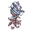
| Entry | Database: PDB / ID: 2q2m | ||||||
|---|---|---|---|---|---|---|---|
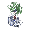
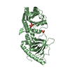
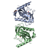
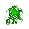
| Title | Beta-lactoglobulin (native) | ||||||
 Components Components | Beta-lactoglobulin | ||||||
 Keywords Keywords | TRANSPORT PROTEIN / LIPOCALIN / VARIANT A | ||||||
| Function / homology |  Function and homology information Function and homology informationretinol binding / long-chain fatty acid binding / extracellular region / identical protein binding Similarity search - Function | ||||||
| Biological species |  | ||||||
| Method |  X-RAY DIFFRACTION / MOLECULAR REPLACEMENT / Resolution: 2.1 Å X-RAY DIFFRACTION / MOLECULAR REPLACEMENT / Resolution: 2.1 Å | ||||||
 Authors Authors | Vijayalakshmi, L. / Krishna, R. / Sankaranarayanan, R. / Vijayan, M. | ||||||
 Citation Citation | Journal: Proteins / Year: 2007 Title: An asymmetric dimer of beta-lactoglobulin in a low humidity crystal form-Structural changes that accompany partial dehydration and protein action. Authors: Vijayalakshmi, L. / Krishna, R. / Sankaranarayanan, R. / Vijayan, M. | ||||||
| History |
|
- Structure visualization
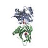
| Structure viewer | Molecule: MolmilJmol/JSmol |
|---|
- Downloads & links
-Download
| PDBx/mmCIF format | 2q2m.cif.gz | 45.7 KB | Display | PDBx/mmCIF format |
|---|---|---|---|---|
| PDB format | pdb2q2m.ent.gz | 32 KB | Display | PDB format |
| PDBx/mmJSON format | 2q2m.json.gz | Tree view | PDBx/mmJSON format | |
| Others |  Other downloads Other downloads |
-Validation report
| Arichive directory | https://data.pdbj.org/pub/pdb/validation_reports/q2/2q2mftp://data.pdbj.org/pub/pdb/validation_reports/q2/2q2m | HTTPS FTP |
|---|
-Related structure data
| Related structure data |  2q2pC  2q39C  1b8eS S: Starting model for refinement C: citing same article ( |
|---|---|
| Similar structure data |


-Links
 PDBj
PDBj

- Assembly
| Deposited unit | 
| ||||||||
|---|---|---|---|---|---|---|---|---|---|
| 1 | 
| ||||||||
| Unit cell |
| ||||||||
| Components on special symmetry positions |
| ||||||||
| Details | The second part of the biologcal assenbly is generated by the twold axis 2-x,y,1/2-z |
-Components
| #1: Protein | Mass: 18387.264 Da / Num. of mol.: 1 / Source method: isolated from a natural source Details: VARIANT A DIFFERS IN A PRIMARY AMINO ACID SEQUENCE FROM VARIANT B AT POSITIONS 64 (ASP-->GLY) AND 118 (VAL-->ALA) Source: (natural) |
|---|---|
| #2: Water | ChemComp-HOH /  Mass: 18.015 Da / Num. of mol.: 137 / Source method: isolated from a natural source / Formula: H2O Mass: 18.015 Da / Num. of mol.: 137 / Source method: isolated from a natural source / Formula: H2O |
| Has protein modification | Y |
-Experimental details
-Experiment
| Experiment | Method: X-RAY DIFFRACTION / Number of used crystals: 1 |
|---|
- Sample preparation
| Crystal | Density Matthews: 2.07 Å3/Da / Density % sol: 40.68 % |
|---|---|
| Crystal grow | Temperature: 293 K / Method: vapor diffusion, hanging drop / pH: 7.4 Details: 0.01M hepes, 2.2 to 2.8 M Ammonium sulphate, NAOH, pH 7.40, VAPOR DIFFUSION, HANGING DROP, temperature 293K |
-Data collection
| Diffraction | Mean temperature: 293 K |
|---|---|
| Diffraction source | Source: ROTATING ANODE / Type: RIGAKU RU200 / Wavelength: 1.5418 |
| Detector | Type: MARRESEARCH / Detector: IMAGE PLATE / Date: May 11, 2004 |
| Radiation | Protocol: SINGLE WAVELENGTH / Monochromatic (M) / Laue (L): M / Scattering type: x-ray |
| Radiation wavelength | Wavelength: 1.5418 Å / Relative weight: 1 |
| Reflection | Resolution: 2.1→19 Å / Num. obs: 8893 / % possible obs: 96.4 % / Observed criterion σ(I): 0 / Biso Wilson estimate: 16.4 Å2 / Rmerge(I) obs: 0.11 / Net I/σ(I): 9.9 |
| Reflection shell | Resolution: 2.1→2.23 Å / Rmerge(I) obs: 0.39 / Mean I/σ(I) obs: 3.7 / % possible all: 97.7 |
- Processing
| Software |
| ||||||||||||||||||||
|---|---|---|---|---|---|---|---|---|---|---|---|---|---|---|---|---|---|---|---|---|---|
| Refinement | Method to determine structure: MOLECULAR REPLACEMENT Starting model: PDB entry 1B8E Resolution: 2.1→19 Å / Rfactor Rfree error: 0.009 / Data cutoff high absF: 1409038.93 / Data cutoff low absF: 0 / Isotropic thermal model: GROUP / Cross valid method: THROUGHOUT / σ(F): 0
| ||||||||||||||||||||
| Solvent computation | Solvent model: FLAT MODEL / Bsol: 73.2231 Å2 / ksol: 0.293624 e/Å3 | ||||||||||||||||||||
| Displacement parameters | Biso mean: 40.4 Å2
| ||||||||||||||||||||
| Refine analyze |
| ||||||||||||||||||||
| Refinement step | Cycle: LAST / Resolution: 2.1→19 Å
| ||||||||||||||||||||
| Refine LS restraints |
| ||||||||||||||||||||
| LS refinement shell | Resolution: 2.1→2.23 Å / Rfactor Rfree error: 0.023 / Total num. of bins used: 6
| ||||||||||||||||||||
| Xplor file |
|
