 Movie
Movie Controller

[English] 日本語
 Yorodumi
- PDB-6ghh: Thermodynamic, Crystallographic and Computational Studies of Non ... -
+ Open data

- Basic information
| Entry | Database: PDB / ID: 6ghh | ||||||
|---|---|---|---|---|---|---|---|
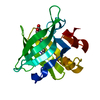
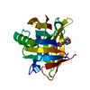
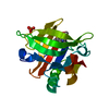
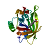
| Title | Thermodynamic, Crystallographic and Computational Studies of Non Mammalian Fatty Acid Binding to Bovine b-Lactoglobulin | ||||||
 Components Components | Beta-lactoglobulin | ||||||
 Keywords Keywords | TRANSPORT PROTEIN / lactoglobulin / fatty acid / Non-Mammalian | ||||||
| Function / homology |  Function and homology information Function and homology informationretinol binding / long-chain fatty acid binding / extracellular region / identical protein binding Similarity search - Function | ||||||
| Biological species |  | ||||||
| Method |  X-RAY DIFFRACTION / MOLECULAR REPLACEMENT / Resolution: 1.9 Å X-RAY DIFFRACTION / MOLECULAR REPLACEMENT / Resolution: 1.9 Å | ||||||
 Authors Authors | Kontopidis, G. / Rovoli, M. | ||||||
 Citation Citation | Journal: Int. J. Biol. Macromol. / Year: 2018 Title: Thermodynamic, crystallographic and computational studies of non-mammalian fatty acid binding to bovine beta-Lactoglobulin. Authors: Rovoli, M. / Thireou, T. / Choiset, Y. / Haertle, T. / Sawyer, L. / Eliopoulos, E. / Kontopidis, G. | ||||||
| History |
|
- Structure visualization
| Structure viewer | Molecule: MolmilJmol/JSmol |
|---|
- Downloads & links
-Download
| PDBx/mmCIF format | 6ghh.cif.gz | 82.7 KB | Display | PDBx/mmCIF format |
|---|---|---|---|---|
| PDB format | pdb6ghh.ent.gz | 62 KB | Display | PDB format |
| PDBx/mmJSON format | 6ghh.json.gz | Tree view | PDBx/mmJSON format | |
| Others |  Other downloads Other downloads |
-Validation report
| Arichive directory | https://data.pdbj.org/pub/pdb/validation_reports/gh/6ghhftp://data.pdbj.org/pub/pdb/validation_reports/gh/6ghh | HTTPS FTP |
|---|
-Related structure data
| Related structure data | 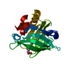 6ge7C 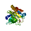 6gf9C  6gfsC 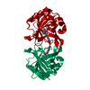 1gx9S S: Starting model for refinement C: citing same article ( |
|---|---|
| Similar structure data |


-Links
 PDBj
PDBj

- Assembly
| Deposited unit | 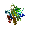
| ||||||||
|---|---|---|---|---|---|---|---|---|---|
| 1 |
| ||||||||
| Unit cell |
|
-Components
| #1: Protein | Mass: 18301.174 Da / Num. of mol.: 1 Source method: isolated from a genetically manipulated source Source: (gene. exp.) |
|---|---|
| #2: Chemical | ChemComp-TDA /   Mass: 214.344 Da / Num. of mol.: 1 / Source method: obtained synthetically / Formula: C13H26O2 / Feature type: SUBJECT OF INVESTIGATION Mass: 214.344 Da / Num. of mol.: 1 / Source method: obtained synthetically / Formula: C13H26O2 / Feature type: SUBJECT OF INVESTIGATION |
| #3: Water | ChemComp-HOH /  Mass: 18.015 Da / Num. of mol.: 76 / Source method: isolated from a natural source / Formula: H2O Mass: 18.015 Da / Num. of mol.: 76 / Source method: isolated from a natural source / Formula: H2O |
| Has protein modification | Y |
-Experimental details
-Experiment
| Experiment | Method: X-RAY DIFFRACTION / Number of used crystals: 1 |
|---|
- Sample preparation
| Crystal | Density Matthews: 2.495 Å3/Da / Density % sol: 50.83 % |
|---|---|
| Crystal grow | Temperature: 293.2 K / Method: vapor diffusion, hanging drop / pH: 7.3 Details: protein 20 mg/mL, ligand 10mM, 20mM Tris buffer, pH 8, which were mixed with 4 microliter of well solution. Drops were equilibrated against 1 mL of well solution containing 1.43 M sodium ...Details: protein 20 mg/mL, ligand 10mM, 20mM Tris buffer, pH 8, which were mixed with 4 microliter of well solution. Drops were equilibrated against 1 mL of well solution containing 1.43 M sodium citrate and 0.1M Hepes, pH 7.5 |
-Data collection
| Diffraction | Mean temperature: 100 K |
|---|---|
| Diffraction source | Source: SEALED TUBE / Type: OXFORD DIFFRACTION NOVA / Wavelength: 1.54 Å |
| Detector | Type: AGILENT ATLAS CCD / Detector: CCD / Date: May 19, 2016 |
| Radiation | Protocol: SINGLE WAVELENGTH / Monochromatic (M) / Laue (L): M / Scattering type: x-ray |
| Radiation wavelength | Wavelength: 1.54 Å / Relative weight: 1 |
| Reflection | Resolution: 1.9→13.8 Å / Num. obs: 17826 / % possible obs: 99.7 % / Redundancy: 4.5 % / Net I/σ(I): 8.2 |
| Reflection shell | Resolution: 1.9→2.01 Å |
- Processing
| Software |
| ||||||||||||||||||||||||||||||||||||||||||||||||||||||||||||||||||||||||||||||||||||||||||||||||||||||||||||||||||||||||||||||||||||||||||||||||||||||||||||||||||||||||||||||||||||||
|---|---|---|---|---|---|---|---|---|---|---|---|---|---|---|---|---|---|---|---|---|---|---|---|---|---|---|---|---|---|---|---|---|---|---|---|---|---|---|---|---|---|---|---|---|---|---|---|---|---|---|---|---|---|---|---|---|---|---|---|---|---|---|---|---|---|---|---|---|---|---|---|---|---|---|---|---|---|---|---|---|---|---|---|---|---|---|---|---|---|---|---|---|---|---|---|---|---|---|---|---|---|---|---|---|---|---|---|---|---|---|---|---|---|---|---|---|---|---|---|---|---|---|---|---|---|---|---|---|---|---|---|---|---|---|---|---|---|---|---|---|---|---|---|---|---|---|---|---|---|---|---|---|---|---|---|---|---|---|---|---|---|---|---|---|---|---|---|---|---|---|---|---|---|---|---|---|---|---|---|---|---|---|---|
| Refinement | Method to determine structure: MOLECULAR REPLACEMENT Starting model: 1GX9 Resolution: 1.9→13.8 Å / Cor.coef. Fo:Fc: 0.962 / Cor.coef. Fo:Fc free: 0.933 / SU B: 9.441 / SU ML: 0.133 / Cross valid method: THROUGHOUT / ESU R: 0.149 / ESU R Free: 0.147 / Details: HYDROGENS HAVE BEEN ADDED IN THE RIDING POSITIONS
| ||||||||||||||||||||||||||||||||||||||||||||||||||||||||||||||||||||||||||||||||||||||||||||||||||||||||||||||||||||||||||||||||||||||||||||||||||||||||||||||||||||||||||||||||||||||
| Solvent computation | Ion probe radii: 0.8 Å / Shrinkage radii: 0.8 Å / VDW probe radii: 1.1 Å | ||||||||||||||||||||||||||||||||||||||||||||||||||||||||||||||||||||||||||||||||||||||||||||||||||||||||||||||||||||||||||||||||||||||||||||||||||||||||||||||||||||||||||||||||||||||
| Displacement parameters | Biso mean: 45.948 Å2
| ||||||||||||||||||||||||||||||||||||||||||||||||||||||||||||||||||||||||||||||||||||||||||||||||||||||||||||||||||||||||||||||||||||||||||||||||||||||||||||||||||||||||||||||||||||||
| Refinement step | Cycle: 1 / Resolution: 1.9→13.8 Å
| ||||||||||||||||||||||||||||||||||||||||||||||||||||||||||||||||||||||||||||||||||||||||||||||||||||||||||||||||||||||||||||||||||||||||||||||||||||||||||||||||||||||||||||||||||||||
| Refine LS restraints |
|