 Movie
Movie Controller

[English] 日本語
 Yorodumi
- PDB-3epy: Crystal Structure of human acyl-CoA binding domain 7 complexed wi... -
+ Open data

- Basic information
| Entry | Database: PDB / ID: 3epy | ||||||
|---|---|---|---|---|---|---|---|
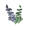
| Title | Crystal Structure of human acyl-CoA binding domain 7 complexed with palmitoyl-Coa | ||||||
 Components Components | Acyl-CoA-binding domain-containing protein 7 | ||||||
 Keywords Keywords | LIPID BINDING PROTEIN / ACYL-COA BINDING PROTEIN / FATTY ACID / LIPID METABOLISM / Structural Genomics / Structural Genomics Consortium / SGC | ||||||
| Function / homology |  Function and homology information Function and homology informationfatty-acyl-CoA binding / Mitochondrial Fatty Acid Beta-Oxidation / fatty acid metabolic process Similarity search - Function | ||||||
| Biological species |  Homo sapiens (human) Homo sapiens (human) | ||||||
| Method |  X-RAY DIFFRACTION / SYNCHROTRON / MOLECULAR REPLACEMENT / Resolution: 2.005 Å X-RAY DIFFRACTION / SYNCHROTRON / MOLECULAR REPLACEMENT / Resolution: 2.005 Å | ||||||
 Authors Authors | Kavanagh, K.L. / Salah, E. / Yue, W.W. / Savitsky, P. / Murray, J.W. / Arrowsmith, C.H. / Weigelt, J. / Edwards, A.M. / Bountra, C. / von Delft, F. ...Kavanagh, K.L. / Salah, E. / Yue, W.W. / Savitsky, P. / Murray, J.W. / Arrowsmith, C.H. / Weigelt, J. / Edwards, A.M. / Bountra, C. / von Delft, F. / Oppermann, U. / Structural Genomics Consortium (SGC) | ||||||
 Citation Citation | Journal: To be Published Title: Crystal Structure of human acyl-CoA binding domain 7 complexed with palmitoyl-Coa Authors: Kavanagh, K.L. / Salah, E. / Yue, W.W. / Savitsky, P. / Murray, J.W. / Arrowsmith, C.H. / Weigelt, J. / Edwards, A.M. / Bountra, C. / von Delft, F. / Oppermann, U. | ||||||
| History |
|
- Structure visualization
| Structure viewer | Molecule: MolmilJmol/JSmol |
|---|
- Downloads & links
-Download
| PDBx/mmCIF format | 3epy.cif.gz | 88.9 KB | Display | PDBx/mmCIF format |
|---|---|---|---|---|
| PDB format | pdb3epy.ent.gz | 67.3 KB | Display | PDB format |
| PDBx/mmJSON format | 3epy.json.gz | Tree view | PDBx/mmJSON format | |
| Others |  Other downloads Other downloads |
-Validation report
| Arichive directory | https://data.pdbj.org/pub/pdb/validation_reports/ep/3epyftp://data.pdbj.org/pub/pdb/validation_reports/ep/3epy | HTTPS FTP |
|---|
-Related structure data
| Related structure data | 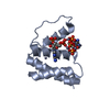 2cb8S S: Starting model for refinement |
|---|---|
| Similar structure data |
-Links
 PDBj
PDBj


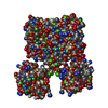
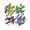
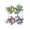
- Assembly
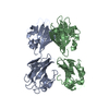
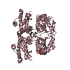
| Deposited unit | 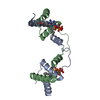
| ||||||||||||||||||||||||||||||||||||||||||||||||||||||||||||||||||||||||||||||||||||||||||||||||||||||||||||||||||||||
|---|---|---|---|---|---|---|---|---|---|---|---|---|---|---|---|---|---|---|---|---|---|---|---|---|---|---|---|---|---|---|---|---|---|---|---|---|---|---|---|---|---|---|---|---|---|---|---|---|---|---|---|---|---|---|---|---|---|---|---|---|---|---|---|---|---|---|---|---|---|---|---|---|---|---|---|---|---|---|---|---|---|---|---|---|---|---|---|---|---|---|---|---|---|---|---|---|---|---|---|---|---|---|---|---|---|---|---|---|---|---|---|---|---|---|---|---|---|---|---|
| 1 | 
| ||||||||||||||||||||||||||||||||||||||||||||||||||||||||||||||||||||||||||||||||||||||||||||||||||||||||||||||||||||||
| 2 |
| ||||||||||||||||||||||||||||||||||||||||||||||||||||||||||||||||||||||||||||||||||||||||||||||||||||||||||||||||||||||
| Unit cell |
| ||||||||||||||||||||||||||||||||||||||||||||||||||||||||||||||||||||||||||||||||||||||||||||||||||||||||||||||||||||||
| Noncrystallographic symmetry (NCS) | NCS domain:
NCS domain segments:
|