 Movie
Movie Controller

[English] 日本語
 Yorodumi
- PDB-1kyq: Met8p: A bifunctional NAD-dependent dehydrogenase and ferrochelat... -
+ Open data

- Basic information
| Entry | Database: PDB / ID: 1kyq | ||||||
|---|---|---|---|---|---|---|---|
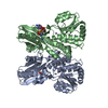
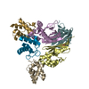
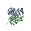
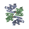
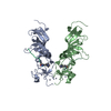
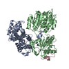
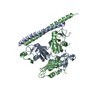
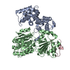
| Title | Met8p: A bifunctional NAD-dependent dehydrogenase and ferrochelatase involved in siroheme synthesis. | ||||||
 Components Components | Siroheme biosynthesis protein MET8 | ||||||
 Keywords Keywords | OXIDOREDUCTASE / LYASE / homodimer | ||||||
| Function / homology |  Function and homology information Function and homology informationprecorrin-2 dehydrogenase / precorrin-2 dehydrogenase activity / sirohydrochlorin ferrochelatase / sirohydrochlorin ferrochelatase activity / siroheme biosynthetic process / sulfate assimilation / protoporphyrin ferrochelatase activity Similarity search - Function | ||||||
| Biological species |  | ||||||
| Method |  X-RAY DIFFRACTION / SYNCHROTRON / MAD / Resolution: 2.2 Å X-RAY DIFFRACTION / SYNCHROTRON / MAD / Resolution: 2.2 Å | ||||||
 Authors Authors | Schubert, H.L. / Raux, E. / Brindley, A.A. / Wilson, K.S. / Hill, C.P. / Warren, M.J. | ||||||
 Citation Citation | Journal: EMBO J. / Year: 2002 Title: The structure of Saccharomyces cerevisiae Met8p, a bifunctional dehydrogenase and ferrochelatase. Authors: Schubert, H.L. / Raux, E. / Brindley, A.A. / Leech, H.K. / Wilson, K.S. / Hill, C.P. / Warren, M.J. #1: Journal: Biophys.J. / Year: 1999Title: The Role of Saccharomyces cerevisiae Met1p and Met8p in Sirohaem and Cobalamin Biosynthesis Authors: Raux, E. / McVeigh, T. / Peters, S.E. / Leustek, T. / Warren, M.J. | ||||||
| History |
| ||||||
| Remark 999 | SEQUENCE During several rounds of independent PCR and sequencing from S. cervisiae, the authors ...SEQUENCE During several rounds of independent PCR and sequencing from S. cervisiae, the authors discovered several discrepancies in the primary sequence: K15R, I33(MSE) (a helpful mutation for solving the structure), E61K, D102N. |
- Structure visualization
| Structure viewer | Molecule: MolmilJmol/JSmol |
|---|
- Downloads & links
-Download
| PDBx/mmCIF format | 1kyq.cif.gz | 191.6 KB | Display | PDBx/mmCIF format |
|---|---|---|---|---|
| PDB format | pdb1kyq.ent.gz | 152.2 KB | Display | PDB format |
| PDBx/mmJSON format | 1kyq.json.gz | Tree view | PDBx/mmJSON format | |
| Others |  Other downloads Other downloads |
-Validation report
| Arichive directory | https://data.pdbj.org/pub/pdb/validation_reports/ky/1kyqftp://data.pdbj.org/pub/pdb/validation_reports/ky/1kyq | HTTPS FTP |
|---|
-Related structure data
| Similar structure data |
|---|


-Links
 PDBj
PDBj- Assembly
| Deposited unit | 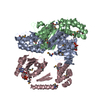
| ||||||||
|---|---|---|---|---|---|---|---|---|---|
| 1 | 
| ||||||||
| 2 | 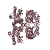
| ||||||||
| Unit cell |
|
-Components
| #1: Protein | Mass: 32289.268 Da / Num. of mol.: 3 Source method: isolated from a genetically manipulated source Source: (gene. exp.) Gene: Met8 / Plasmid: pET14b / Production host:  #2: Chemical |   Mass: 663.425 Da / Num. of mol.: 3 / Source method: obtained synthetically / Formula: C21H27N7O14P2 / Comment: NAD*YM Mass: 663.425 Da / Num. of mol.: 3 / Source method: obtained synthetically / Formula: C21H27N7O14P2 / Comment: NAD*YM#3: Water | ChemComp-HOH / |  Mass: 18.015 Da / Num. of mol.: 614 / Source method: isolated from a natural source / Formula: H2O Mass: 18.015 Da / Num. of mol.: 614 / Source method: isolated from a natural source / Formula: H2OHas protein modification | Y | |
|---|
-Experimental details
-Experiment
| Experiment | Method: X-RAY DIFFRACTION / Number of used crystals: 1 |
|---|
- Sample preparation
| Crystal | Density Matthews: 2.89 Å3/Da / Density % sol: 57.46 % | |||||||||||||||||||||||||||||||||||||||||||||||||
|---|---|---|---|---|---|---|---|---|---|---|---|---|---|---|---|---|---|---|---|---|---|---|---|---|---|---|---|---|---|---|---|---|---|---|---|---|---|---|---|---|---|---|---|---|---|---|---|---|---|---|
| Crystal grow | Temperature: 298 K / Method: vapor diffusion, hanging drop / pH: 8.5 Details: PEG 4000, CaCl2, Tris, pH 8.5, VAPOR DIFFUSION, HANGING DROP, temperature 298K | |||||||||||||||||||||||||||||||||||||||||||||||||
| Crystal | *PLUS Density % sol: 60 % | |||||||||||||||||||||||||||||||||||||||||||||||||
| Crystal grow | *PLUS | |||||||||||||||||||||||||||||||||||||||||||||||||
| Components of the solutions | *PLUS
|
-Data collection
| Diffraction | Mean temperature: 100 K |
|---|---|
| Diffraction source | Source: SYNCHROTRON / Site: SSRL  / Beamline: BL9-1 / Wavelength: 0.97 Å / Beamline: BL9-1 / Wavelength: 0.97 Å |
| Detector | Type: MARRESEARCH / Detector: IMAGE PLATE / Date: Mar 30, 2001 / Details: Flat mirror (vertical focusing) |
| Radiation | Monochromator: single crystal Si(311) bent monochromator (horizontal focusing) Protocol: SINGLE WAVELENGTH / Monochromatic (M) / Laue (L): M / Scattering type: x-ray |
| Radiation wavelength | Wavelength: 0.97 Å / Relative weight: 1 |
| Reflection | Resolution: 1.9→30 Å / Num. all: 64205 / Num. obs: 64205 / % possible obs: 100 % / Observed criterion σ(F): 0 / Observed criterion σ(I): -3 / Redundancy: 6.1 % / Biso Wilson estimate: 28 Å2 / Rmerge(I) obs: 0.067 / Rsym value: 0.067 / Net I/σ(I): 15.7 |
| Reflection shell | Resolution: 2.2→2.28 Å / Redundancy: 2.1 % / Rmerge(I) obs: 0.304 / Mean I/σ(I) obs: 2.9 / Num. unique all: 9417 / Rsym value: 0.304 / % possible all: 100 |
| Reflection | *PLUS Highest resolution: 2.2 Å / Lowest resolution: 20 Å / Num. obs: 56148 / % possible obs: 98 % / Num. measured all: 117894 / Rmerge(I) obs: 0.07 |
| Reflection shell | *PLUS % possible obs: 85.6 % / Rmerge(I) obs: 0.304 / Mean I/σ(I) obs: 3 |
- Processing
| Software |
| ||||||||||||||||||||
|---|---|---|---|---|---|---|---|---|---|---|---|---|---|---|---|---|---|---|---|---|---|
| Refinement | Method to determine structure: MAD / Resolution: 2.2→30 Å / σ(F): 0 / σ(I): 0 Details: REFMAC MKLF. PORTIONS OF THE NAD LIGANDS WERE MISSING IN THE DENSITY.
| ||||||||||||||||||||
| Refinement step | Cycle: LAST / Resolution: 2.2→30 Å
| ||||||||||||||||||||
| Refine LS restraints |
| ||||||||||||||||||||
| Refinement | *PLUS % reflection Rfree: 5 % / Rfactor all: 0.22 / Rfactor obs: 0.221 / Rfactor Rfree: 0.287 / Rfactor Rwork: 0.221 | ||||||||||||||||||||
| Solvent computation | *PLUS | ||||||||||||||||||||
| Displacement parameters | *PLUS | ||||||||||||||||||||
| Refine LS restraints | *PLUS
|
